Figures & data
Table 1. List of commercially available vaccines administered by the mucosal routes*,**
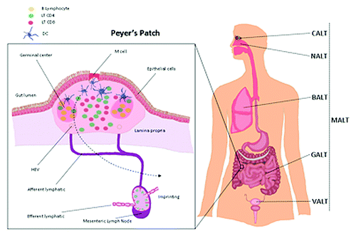
Figure 1. Mucosal associated lymphoid tissue (MALT) are organized lymphoid structures present in surface area in contact with environment such as the lung (bronchus-associated lymphoid tissue (BALT), the nose (Nasal-associated lymphoid tissue (NALT) and the gut (Gut-associated lymphoid tissue (GALT). Peyer’s patch present in the GALT are often presented as a model of the MALT organization. It is located in the lamina propria layer of the small intestine and in the ileum in humans. This lymphoid structure between the lumen of the intestine and the mesenteric lymph node is the place of the priming of a mucosal immune response.