1,468
Views
20
CrossRef citations to date
0
Altmetric
Research Paper

Depletion of tumor-associated macrophages enhances the anti-tumor immunity induced by a Toll-like receptor agonist-conjugated peptide

, , , &
Pages 3241-3250 | Received 06 Mar 2014, Accepted 18 May 2014, Published online: 01 Nov 2014

Figures & data

Figure 1. Pam2IDG enhances BMDC maturation and CTL activities to improve anti-tumor immunity. BMDCs were treated with 1 μM IDG or Pam2IDG at the indicated concentration for 18 h. (A) The level of IL-12p40 in the supernatant was determined by ELISA. (B) The mean fluorescence intensities of the surface molecules were analyzed by flow cytometry. Relative MFI = (MFI of IDG- or Pam2IDG-treated cells / MFI of untreated cells) x 100%. BMDCs cultured from WT, TLR1KO, TLR2KO, and TLR6KO mice were treated with 1 μM IDG, Pam2IDG, or 100 ng/ml LPS at the indicated concentration for 18 h. The levels of (C) IL-12p40 and (D) IL-6 in the supernatant were determined by ELISA.
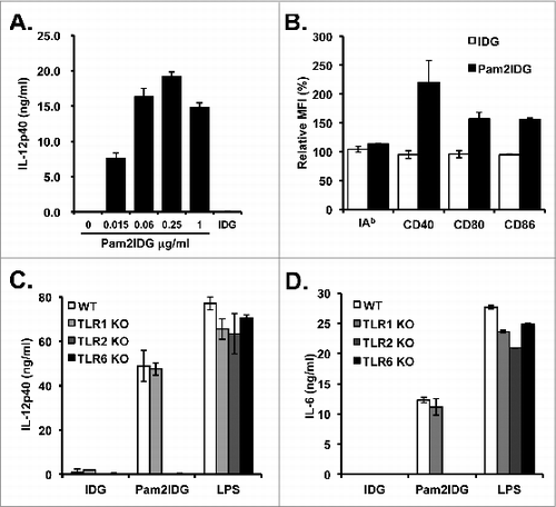
Figure 1. Pam2IDG enhances BMDC maturation and CTL activities to improve anti-tumor immunity. BMDCs were treated with 1 μM IDG or Pam2IDG at the indicated concentration for 18 h. (A) The level of IL-12p40 in the supernatant was determined by ELISA. (B) The mean fluorescence intensities of the surface molecules were analyzed by flow cytometry. Relative MFI = (MFI of IDG- or Pam2IDG-treated cells / MFI of untreated cells) x 100%. BMDCs cultured from WT, TLR1KO, TLR2KO, and TLR6KO mice were treated with 1 μM IDG, Pam2IDG, or 100 ng/ml LPS at the indicated concentration for 18 h. The levels of (C) IL-12p40 and (D) IL-6 in the supernatant were determined by ELISA.

Figure 2. Pam2IDG increases the number of IFN-γ-producing cells. C57BL/6 naïve mice were immunized with 10 μg of IDG or Pam2IDG via the footpad on days 0 and 5. On day 10, the lymphocytes were collected from inguinal lymph nodes. (A) A total of 2 × 10⁵ lymphocytes from each treatment group were re-stimulated with 5 μg/ml RAH or control peptide in anti-IFN-γ antibody-coated plates for 48 h. The IFN-γ-producing cells were examined by ELISPOT. (B) The lymphocytes were double-stained with RAH tetramer (Tet-RAH) and anti-CD8 antibody and analyzed by flow cytometry. The data are presented as the cell numbers of Tet-RAH+ CD8+ T cells from two independent experiments. (C) In vivo analysis of WT, TLR1KO, TLR2KO, and TLR6KO mice immunized as in (A), and the numbers of IFN-γ-producing cells were then examined by ELISPOT. (D) In vitro analysis of WT, TLR1KO, TLR2KO, TLR6KO and MyD88KO BMDCs pulsed with 1 μM IDG or Pam2IDG for 2.5 h (the free peptides were then washed out). A total of 2 × 10⁴ peptide-pulsed BMDCs were co-cultured with 2 × 10⁵ responder cells, which were purified from CD8+ T cells from the spleens of RAH/IFA-immunized mice. After 48 h, the IFN-γ-producing cells were examined by ELISPOT.
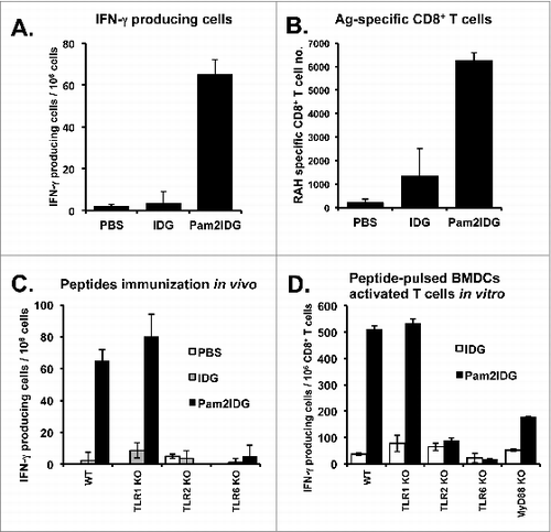
Figure 2. Pam2IDG increases the number of IFN-γ-producing cells. C57BL/6 naïve mice were immunized with 10 μg of IDG or Pam2IDG via the footpad on days 0 and 5. On day 10, the lymphocytes were collected from inguinal lymph nodes. (A) A total of 2 × 10⁵ lymphocytes from each treatment group were re-stimulated with 5 μg/ml RAH or control peptide in anti-IFN-γ antibody-coated plates for 48 h. The IFN-γ-producing cells were examined by ELISPOT. (B) The lymphocytes were double-stained with RAH tetramer (Tet-RAH) and anti-CD8 antibody and analyzed by flow cytometry. The data are presented as the cell numbers of Tet-RAH+ CD8+ T cells from two independent experiments. (C) In vivo analysis of WT, TLR1KO, TLR2KO, and TLR6KO mice immunized as in (A), and the numbers of IFN-γ-producing cells were then examined by ELISPOT. (D) In vitro analysis of WT, TLR1KO, TLR2KO, TLR6KO and MyD88KO BMDCs pulsed with 1 μM IDG or Pam2IDG for 2.5 h (the free peptides were then washed out). A total of 2 × 10⁴ peptide-pulsed BMDCs were co-cultured with 2 × 10⁵ responder cells, which were purified from CD8+ T cells from the spleens of RAH/IFA-immunized mice. After 48 h, the IFN-γ-producing cells were examined by ELISPOT.

Figure 3. Pam2IDG immunization inhibits palpable tumor growth. C57BL/6 naïve mice were inoculated subcutaneously with 2 × 10⁵ TC-1 tumor cells in the right leg. (A) Seven days before tumor inoculation, the mice were subcutaneously immunized with PBS or 30 μg of IDG or Pam2IDG (n = 5 in each group). (B) The TC-1 tumor-bearing mice were immunized subcutaneously with PBS or 30 μg of IDG or Pam2ID on day 7 (n = 5 in each group). (C) The TC-1 tumor-bearing mice were subcutaneously immunized with PBS or 10 μg of IDG or Pam2IDG on days 7 and 14 (n = 6 in each group). (D) The TC-1 tumor-bearing mice were immunized with 30 μg of Pam2IDG on day 14 and 10 μg of Pam2IDG on days 14 and 21 (n = 6 in each group).
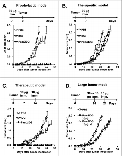
Figure 3. Pam2IDG immunization inhibits palpable tumor growth. C57BL/6 naïve mice were inoculated subcutaneously with 2 × 10⁵ TC-1 tumor cells in the right leg. (A) Seven days before tumor inoculation, the mice were subcutaneously immunized with PBS or 30 μg of IDG or Pam2IDG (n = 5 in each group). (B) The TC-1 tumor-bearing mice were immunized subcutaneously with PBS or 30 μg of IDG or Pam2ID on day 7 (n = 5 in each group). (C) The TC-1 tumor-bearing mice were subcutaneously immunized with PBS or 10 μg of IDG or Pam2IDG on days 7 and 14 (n = 6 in each group). (D) The TC-1 tumor-bearing mice were immunized with 30 μg of Pam2IDG on day 14 and 10 μg of Pam2IDG on days 14 and 21 (n = 6 in each group).

Figure 4. Therapeutic effects of Pam2IDG are improved by the reduction of immunosuppressive factors. C57BL/6 naïve mice were inoculated subcutaneously with 2 × 10⁵ TC-1 tumor cells in the right leg. (A and B) TC-1 tumor-bearing mice were intraperitoneally injected with 1 mg of clodronate/liposome on day 13 and subcutaneously immunized with 30 μg of Pam2IDG on day 14 (n = 10) in each group; Pam2IDG combined with clodronate vs. Pam2IDG, P < 0.01). (C and D) TC-1 tumor-bearing mice were intraperitoneally injected with 200 μg of anti-IL10R antibody on day 13 and subcutaneously immunized with 30 μg of Pam2IDG on day 14 (n = 6 in each group; Pam2IDG combined with anti-IL10R antibody vs. Pam2IDG, P < 0.01). (E and F) TC-1 tumor-bearing mice were intraperitoneally injected with 0.5 mg/kg celecoxib at 2-d intervals until day 30 and subcutaneously immunized with 30 μg of Pam2IDG on day 14 (n = 10 in each group; Pam2IDG combined with celecoxib vs. Pam2IDG, P < 0.01)
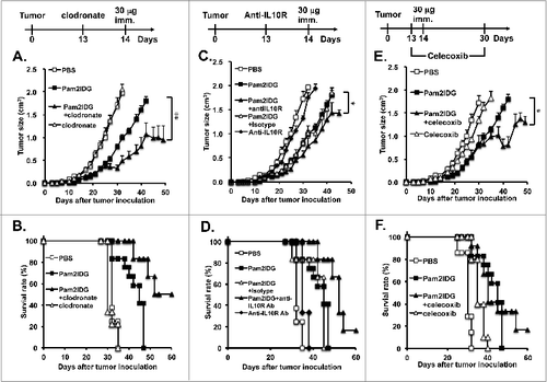
Figure 4. Therapeutic effects of Pam2IDG are improved by the reduction of immunosuppressive factors. C57BL/6 naïve mice were inoculated subcutaneously with 2 × 10⁵ TC-1 tumor cells in the right leg. (A and B) TC-1 tumor-bearing mice were intraperitoneally injected with 1 mg of clodronate/liposome on day 13 and subcutaneously immunized with 30 μg of Pam2IDG on day 14 (n = 10) in each group; Pam2IDG combined with clodronate vs. Pam2IDG, P < 0.01). (C and D) TC-1 tumor-bearing mice were intraperitoneally injected with 200 μg of anti-IL10R antibody on day 13 and subcutaneously immunized with 30 μg of Pam2IDG on day 14 (n = 6 in each group; Pam2IDG combined with anti-IL10R antibody vs. Pam2IDG, P < 0.01). (E and F) TC-1 tumor-bearing mice were intraperitoneally injected with 0.5 mg/kg celecoxib at 2-d intervals until day 30 and subcutaneously immunized with 30 μg of Pam2IDG on day 14 (n = 10 in each group; Pam2IDG combined with celecoxib vs. Pam2IDG, P < 0.01)

Figure 5. Pam2IDG immunization combined with clodronate increases tumor-infiltrating M1/M2 macrophage ratio. Five C57BL/6 naïve mice in each group were subcutaneously inoculated with 2 × 10⁵ TC-1 tumor cells in the right leg. The TC-1 tumor-bearing mice were intraperitoneally injected with 1 mg of clodronate/liposome on day 13 and subcutaneously immunized with or without 30 μg of Pam2IDG on day 14. The tumor infiltrates were analyzed on day 25 by flow cytometry. (A) The TNF-α− and (B) IL10-secreting F4/80+ CD11b+ cells indicate the M1 and M2 macrophages, respectively. (C) The ratio was calculated as the percentage of M1 macrophages divided by the percentage of M2 macrophages. (D) The RAH-specific CTLs were detected using a tetramer. P+C: mice were treated with clodronate and then immunized with Pam2IDG.
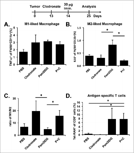
Figure 5. Pam2IDG immunization combined with clodronate increases tumor-infiltrating M1/M2 macrophage ratio. Five C57BL/6 naïve mice in each group were subcutaneously inoculated with 2 × 10⁵ TC-1 tumor cells in the right leg. The TC-1 tumor-bearing mice were intraperitoneally injected with 1 mg of clodronate/liposome on day 13 and subcutaneously immunized with or without 30 μg of Pam2IDG on day 14. The tumor infiltrates were analyzed on day 25 by flow cytometry. (A) The TNF-α− and (B) IL10-secreting F4/80+ CD11b+ cells indicate the M1 and M2 macrophages, respectively. (C) The ratio was calculated as the percentage of M1 macrophages divided by the percentage of M2 macrophages. (D) The RAH-specific CTLs were detected using a tetramer. P+C: mice were treated with clodronate and then immunized with Pam2IDG.