Figures & data
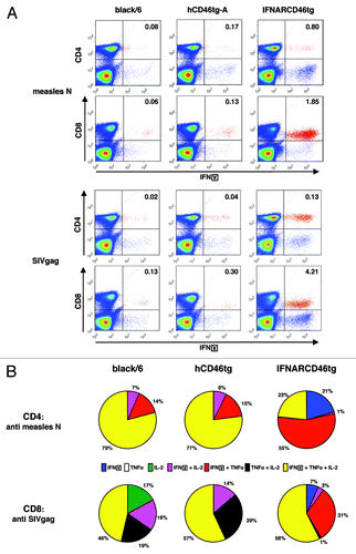
Figure 1. Detection of hCD46 expression on murine PBMCs. Whole blood was isolated from individual mice of five different mouse stains (black/6, hCD46tg-A, hCD46tg-B, hCD46tg-C, and IFNARCD46tg) and stained against hCD46. Median fluorescence intensity was measured by FACS analysis.
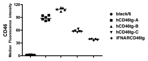
Figure 2. Dose response experiment. A) black/6, hCD46tg-A, and IFNARCD46tg mice were immunized i.m with 103, 104, or 105 pfu rMVb. Anti-measles N humoral immune response was determined by ELISA 4 wk post immunization. ELISA positivity is defined as 3 × over negative control sera.
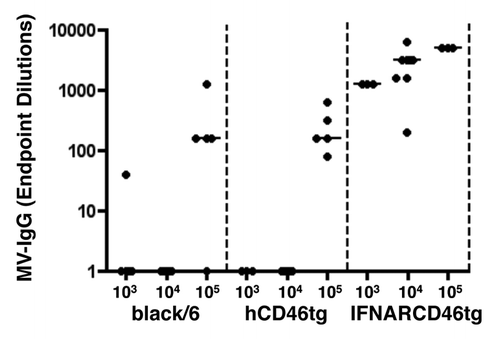
Figure 3. Humoral immune response against rMVb-HIVenv. black/6, hCD46tg-A, and IFNARCD46tg mice were immunized intranasal (i.n) with 105 pfu rMVb2-HIVenv followed by an intramuscular (i.m) boost after 4 wk. Humoral responses against MV-N and HIVenv were assessed 6 wk post immunization.
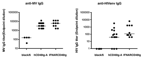
Figure 4. Cellular immune response against rMV-SIVgag: Groups of eight mice of black/6 (black circle), hCD46tg-A (gray triangle), and IFNARCD46tg (gray square) mice were immunized i.m with 105 pfu rMV-SIVgag or 105 pfu UV inactivated rMV-SIVgag (UV-rMV-SIVgag). Cellular immune response was assessed by IFNγ ELISpot 2 wk post immunization. Median values are depicted as line. (A) Spot Forming Cells (SFC) against measles N. (B) SFC against SIVgag. *1 P = 0.003** / *2 P = 0.049* / *3 P = 0.015*
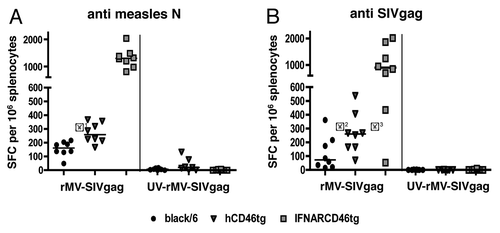
Figure 5. Intracellular cytokine expression profile of rMV-SIVgag induced by CD4+ and CD8+ T-cells against measles N or SIVgag as detected by intracellular cytokine FACS analysis. Five black/6, hCD46tg-A, and IFNARCD46tg mice were immunized with 105 pfu rMVb2-SIVgag. Splenocytes were isolated 2 wk post immunization and restimulated in vitro with either MV-N or SIVgag peptide pools. CD4+ and CD8+ T-cells were stained against IFNγ, TNFα, and IL-2. Negative controls, cultured with media alone, showed less than 0.04% double positive cells in the upper right quadrant when stained against IFNγ and CD4+ or CD8+ (data not shown). (A) Representative FACS results of the three mouse strains. Upper 2 figure lines show the response against MV-N. Lower 2 figure lines show the response against SIVgag. Percentages of IFNγ positive cells are calculated in relation to CD4+ or CD8+ positive cells only. (B) Percentile IFNγ, IL-2, TNFα cytokine distribution for CD4+ T-cells reactive against MV-N and CD8+ T-cells reactive upon SIVgag restimulation.