
Figures & data
Table 1. Biotin binding assay of MAV and SAV
Figure 1. Depiction of self-assembled vaccines structures. (A) Structure of MtbHSP70-avidin (MAV) and biotinylated antigens. (B) Structures of OVA peptides for SAV-OVA studies described in . (C) and (D) Structures of short (C) and long (D) Flu peptides described in for SAV1 and SAV2 used in Flu study.
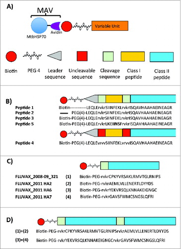
Figure 2. Characterization of Pfēnex’ MtbHSP70-avidin (MAV). (A) 500 nanograms of MAV were electrophoresed on a 4–12% Bis-Tris NuPAGE gel run in MOPS buffer at constant 200 V for 1 hr. (B) Reproduction of the capillary gel electrophoresis (CGE) profile provided by Pfēnex. 150 nl of purified MAV (976.2 ng/nl) were injected into the microfluidic channel of the LabChip GX/II using electrokinetic injection. Applying a voltage across the channel effected protein separation. Protein bands were detected via laser induced fluorescence. Arrows indicate the migration position of MtbHSP70-avidin. (C) MtbHSP70-avidin ATPase activity assessment. A series of MtbHSP70-avidin concentrations (25 – 500 ng) were incubated as described in materials and methods. (D, E, F and G) Flow cytometric analysis of splenocytes and lymph node cells from ovalbumin immunized mice as described in Materials and Methods. Splenocytes (D, F) and lymph node cells (E, G) were prepared as described in Materials and Methods and stimulated with medium alone, SIINFEKL (D, E; 10 μg/ml) or ISQAVHAAHAEINEAGR (F, G; 10 μg/ml) for 24 hours at 37°C. The percent of CD3+CD8+IFN-γ+ and CD3+CD4+IFN-γ+ cells above medium alone was determined and plotted as percent above control. (H) Gel-shift assay of SAV1 and SAV2. 400 to 800 ng of protein were denatured and subjected to electrophoresis on a Novex® 4–12% Bis-Tris NuPAGE gel run in MOPS buffer at constant 200 V for 5 hr. Arrows indicate MAV, SAV1 and SAV2. (I) SAV1 and SAV2 ATPase activity. Three different concentrations of MAV, SAV1 and SAV2 were assayed for their ability to hydrolyze ATP as described in Materials and Methods. Abbreviations: MAV - Mycobacterium tuberculosis heat shock protein 70 fused to avidin; OVA - ovalbumin; PEG - polyethylene glycol; SAV1 - MAV assembled with flu specific peptides 1–4 (); SAV2 - MAV assembled with flu specific peptides 5 and 6 ().
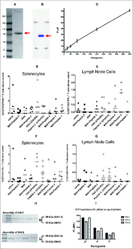
Table 2. Epitopes and peptides sequences used for OVA, Flu and Lassa fever virus vaccines
Table 3. Immunization schemes
Table 4. Antibody responses against immunizing components of the vaccines*
Figure 3. Flow cytometric analysis of splenocytes and lymphocytes from FluVax, SAV1 and SAV2 immunized mice. Splenocytes and lymphocytes were prepared as described in Materials and Methods and stimulated with the indicated flu peptides (10 μg/ml) or medium alone for 24 hours at 37°C. Brefeldin A was added 4 hours prior to harvest to inhibit protein transport. Cells were stained for CD3, CD4, CD8, IL-4 and interferon-γ fixed and analyzed on a BD LSR FortessaTM. The percent of CD3+CD4+IFN-γ+ cells above medium alone was determined and plotted as percent above control. (A) Splenocytes response, (B) lymph node cells response. The percent of CD3+CD4+IL-4+ cells above medium alone was determined and plotted as percent above control for (C) splenocytes response, (D) lymph node cells response. Numbers in parenthesis indicate non-responders.
Figure 4. Structure and sequences of Lassa fever virus immunogenic peptides. (A) Basic framework for the synthesis of immunogenic concatenated class I and class II peptides. (B) Biotinylated Lassa fever virus specific peptides.
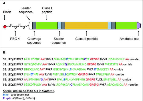
Figure 5. Characterization of the self-assembled Lassa fever virus vaccine (SAVL). A) ATPase activity of MAV, MAVf, and SAVL using 25, 50 and 100 ng. The assay was carried out as described in Materials and Methods. (B) Flow cytometric analysis of splenocytes from mice immunized with SAVL. Splenocytes from mice immunized with PBS, peptides, MAVf and SAVL were prepared as described and stimulated with medium alone or a pool of class II peptides (10 μg/ml). Total incubation time was 24 hours at 37°C. Brefeldin A was added 4 hours prior to harvest. (C) Comparison of splenocytes response to class II Lassa fever virus specific peptides between different immunization groups.
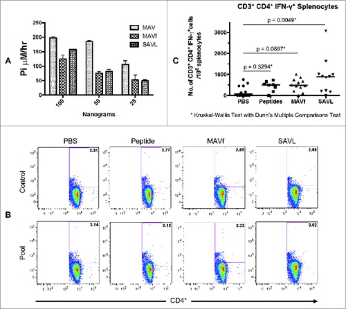
Figure 6. VaxCelerate time line.
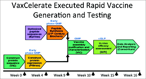
Table 5. Comparison of T cell immune responses observed in this study with other EID vaccines