610
Views
6
CrossRef citations to date
0
Altmetric
Research Papers

Comparison of virulence between the Yersinia pestis Microtus 201, an avirulent strain to humans, and the vaccine strain EV in rhesus macaques, Macaca mulatta

, , , , , , , , , , , , , , & show all
Pages 3552-3560 | Received 31 May 2014, Accepted 13 Aug 2014, Published online: 10 Feb 2015

Figures & data

Figure 1. Post-mortem examination of gross changes in the organs from the animal infected with the Y. pestis Microtus 201 or the vaccine EV. (A and B, a and c) Photos taken from the animals infected intravenously with approximately 109 CFU of the Y. pestis Microtus 201 and the vaccine EV. (A and B, b and d) Photos taken from the animals infected intravenously with approximately 1010 CFU of the Y. pestis Microtus 201 and the vaccine EV.
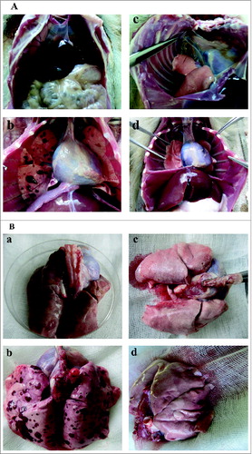
Figure 1. Post-mortem examination of gross changes in the organs from the animal infected with the Y. pestis Microtus 201 or the vaccine EV. (A and B, a and c) Photos taken from the animals infected intravenously with approximately 109 CFU of the Y. pestis Microtus 201 and the vaccine EV. (A and B, b and d) Photos taken from the animals infected intravenously with approximately 1010 CFU of the Y. pestis Microtus 201 and the vaccine EV.

Table 1. Microbiological examination of tissue bacterial load in the Chinese-origin rhesus macaques following Y. pestis infection

Figure 2. The F1 antigen of Y. pestis was identified by immunohistochemitry staining. Bacteria were visualized as those expressing F1 antigen (brown and yellow stain). (A) Tissue sections from the animals infected intravenously with 9 × 10⁸ CFU of the Y. pestis Microtus 201. (B) Tissue sections from the animals infected intravenously with 1.28 × 10¹⁰ CFU of the Y. pestis Microtus 201. (C) Tissue sections from the animals infected intravenously with 9.6 × 10⁸ CFU of the vaccine EV. (D) Tissue sections from the animals infected intravenously with 1.5 × 10¹⁰ CFU of the vaccine EV. (E) Tissue sections from one normal animal that was neither immunized with the Y. pestis Microtus 201 or the EV vaccine. Numerous bacteria were observed in the tissue of hearts (a), livers (b), spleens (c), lungs (d), kidneys (e) and lymph nodes (f) of the dead animals after infection with approximately 1010 CFU of the Y. pestis Microtus 201 (B). No bacterium was found in other groups of animals (A, C and D) and the control animal (E).
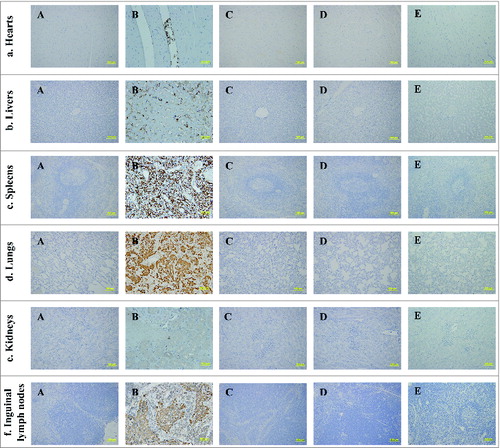
Figure 2. The F1 antigen of Y. pestis was identified by immunohistochemitry staining. Bacteria were visualized as those expressing F1 antigen (brown and yellow stain). (A) Tissue sections from the animals infected intravenously with 9 × 10⁸ CFU of the Y. pestis Microtus 201. (B) Tissue sections from the animals infected intravenously with 1.28 × 10¹⁰ CFU of the Y. pestis Microtus 201. (C) Tissue sections from the animals infected intravenously with 9.6 × 10⁸ CFU of the vaccine EV. (D) Tissue sections from the animals infected intravenously with 1.5 × 10¹⁰ CFU of the vaccine EV. (E) Tissue sections from one normal animal that was neither immunized with the Y. pestis Microtus 201 or the EV vaccine. Numerous bacteria were observed in the tissue of hearts (a), livers (b), spleens (c), lungs (d), kidneys (e) and lymph nodes (f) of the dead animals after infection with approximately 1010 CFU of the Y. pestis Microtus 201 (B). No bacterium was found in other groups of animals (A, C and D) and the control animal (E).

Figure 3. Survival analysis of the Chinese-origin rhesus macaques after intravenous infection with approximately 1010 CFU of the Y. pestis Microtus 201 and the vaccine EV.
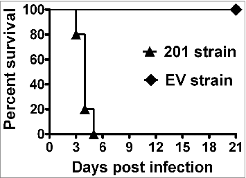
Figure 3. Survival analysis of the Chinese-origin rhesus macaques after intravenous infection with approximately 1010 CFU of the Y. pestis Microtus 201 and the vaccine EV.

Figure 4. Histopathologic observation of the tissues from the animals infected with the Y. pestis Microtus 201 and the vaccine EV. Tissue sections were stained with hematoxylin and eosin for pathological examination after infection with Y. pestis. (A) Tissue sections from the animals infected intravenously with 9 × 10⁸ CFU of the Y. pestis Microtus 201. (B) Tissue sections from the animals infected intravenously with 1.28 × 10¹⁰ CFU of the Y. pestis Microtus 201. (C) Tissue sections from the animals infected intravenously with 9.6 × 10⁸ CFU of the vaccine EV. (D) Tissue sections from the animals infected intravenously with 1.5 × 10¹⁰ CFU of the vaccine EV. (E) Tissue sections from one normal animal.
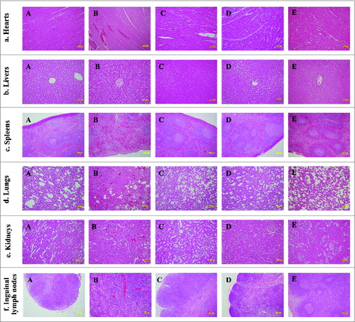
Figure 4. Histopathologic observation of the tissues from the animals infected with the Y. pestis Microtus 201 and the vaccine EV. Tissue sections were stained with hematoxylin and eosin for pathological examination after infection with Y. pestis. (A) Tissue sections from the animals infected intravenously with 9 × 10⁸ CFU of the Y. pestis Microtus 201. (B) Tissue sections from the animals infected intravenously with 1.28 × 10¹⁰ CFU of the Y. pestis Microtus 201. (C) Tissue sections from the animals infected intravenously with 9.6 × 10⁸ CFU of the vaccine EV. (D) Tissue sections from the animals infected intravenously with 1.5 × 10¹⁰ CFU of the vaccine EV. (E) Tissue sections from one normal animal.

Reprints and Corporate Permissions

Please note: Selecting permissions does not provide access to the full text of the article, please see our help page How do I view content?

To request a reprint or corporate permissions for this article, please click on the relevant link below:

Academic Permissions

Please note: Selecting permissions does not provide access to the full text of the article, please see our help page How do I view content?

Obtain permissions instantly via Rightslink by clicking on the button below:

If you are unable to obtain permissions via Rightslink, please complete and submit this Permissions form. For more information, please visit our Permissions help page.