Figures & data
Figure 1. In vivo study design.
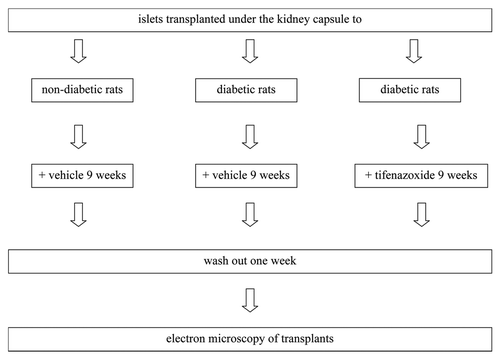
Figure 2. Blood glucose (A) and body weight (B) of transplanted rats that were non-diabetic and vehicle-treated, diabetic and vehicle-treated or diabetic and tifenazoxide-treated. There were eight rats in each group. Treatment was removed after 9 weeks i.e., after day 63.
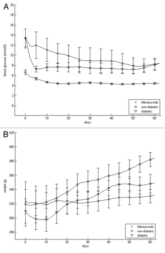
Table 1. Morphometric parameters of islets cell mitochondria
Figure 3. Quantitative morphology of primary (upper panel) and secondary (lower panel) mitochondrial parameters. Data were collected from 22 sections per islet graft. There were eight transplants per group. Totally 528 EM sections were evaluated (176 from each treatment group i.e., approx. 25,000 mitochondria altogether). *p < 0.05 vs. mitochondria from transplants to non-diabetic animals, †p < 0.05 vs. mitochondria from transplants to vehicle-treated diabetic animals. NN414 = tifenazoxide.
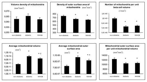
Figure 4. Electron microscopy showing representative sections of β-cells from transplanted islets to non-diabetic, vehicle-treated (A), diabetic, vehicle-treated (B and D) and diabetic, tifenazoxide-treated (C) rats. (E) shows an example of a likely fusion event in a section from a diabetic vehicle-treated rat. In (B) black arrows = mitochondria. In (C) black arrows = mature granule and white arrows = immature granule.
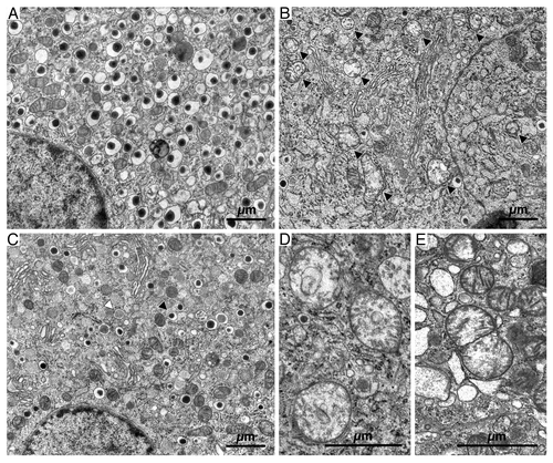
Figure 5. Frequency distribution of average mitochondrial volume (A) and average mitochondrial outer surface area (B).
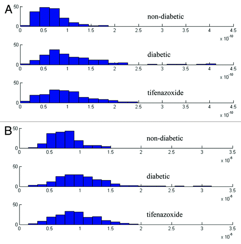
Figure 6. Quantitative morphology of primary (upper panel) and secondary (lower panel) mitochondrial parameters of islets cultured in vitro. Data were collected from 22 sections per rat. Data from 2 weeks (n = 2) and 3 weeks (n = 1) culture in 11 or 27 mmol/l glucose were pooled since quantitative morphology was similar after 2 and 3 weeks culture. *p < 0.05 vs. mitochondria from islets cultured in 11 mmol/l glucose.
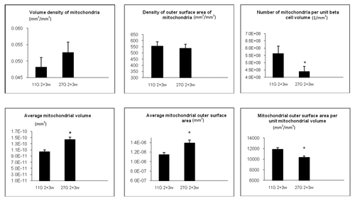
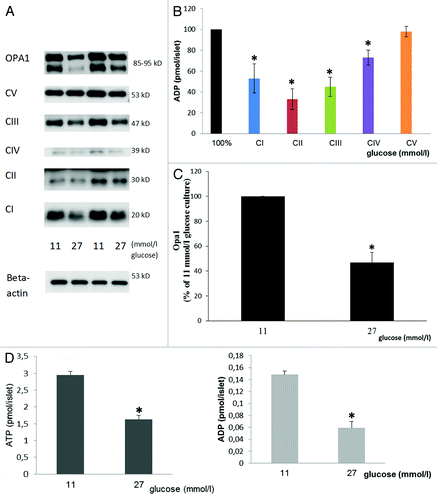
Figure 7. Effects of 3 weeks culture in vitro in 11 or 27 mmol/l glucose on sub-units of mitochondrial complexes I-V (A and quantified in B), OPA 1 (A and quantified in C) and on ATP and ADP levels (D). Immunoblotting was performed on islets cultured either at 27 or 11 mmol/l glucose. In (A), (B) and (C) mean ± SEM of four individual experiments expressed as percentage of results in islets cultured in 11 mmol/l glucose (100%). *p < 0.05 or less vs. 11 mmol/l glucose culture. ATP and ADP levels: Mean ± SEM of four individual experiments. *p < 0.001 or less vs. 11 mmol/l glucose culture.