Figures & data
Figure 1. Manganese is uptaken by human islets. T1-weighted magnetic resonance images of pellets of human islets from non-diabetic multi-organ donors before (A) and after a 30 min exposure to MnCl2 (B). C) Manganese significantly enhanced the MRI signal to noise ratio (16.0 ± 1.3 vs. 41.6 ± 0.7, p = 0.0003). Data are mean + SEM signal-to-noise ratio.
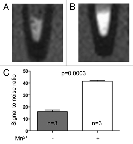
Table 1. MRI indication
Figure 2. The MRI signal enhancement of pancreas is enhanced by manganese. T1-weighted magnetic resonance imaging showing the pancreas (area limited by the dashed line) before (A, C) and 20 min after Mn-DPDP infusion (B, D) of a normoglycemic (A, B) and a type 2 diabetic patient (C, D). In both patients, the MRI signal of pancreas was enhanced by the manganese infusion. E) This enhancement was significantly higher in normoglycemic than in type 2 diabetic patients. Data are mean + SEM signal enhancement, expressed as % of the signal evaluated prior to the manganese infusion.
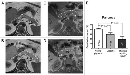
Figure 3. The MEMRI enhancement of pancreas correlated with the BMI of diabetic patients. The pancreas enhancement due to Mn2+ was inversely correlated with the BMI (A), but not the age of the diabetic patients (B), as assessed by Pearson’s correlation.
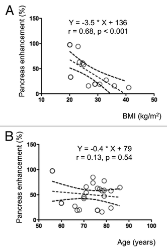
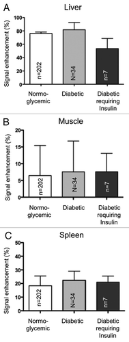
Figure 4. The MEMRI signal of liver (A), muscle (B) and spleen (C) does not differentiate normoglycemic patients from diabetic patients. The signal enhancement of liver (A), muscle (B) and spleen (C) was not statistically different between normoglycemic and type 2 diabetic patients. Data are mean + SEM signal enhancement, expressed as % of the signal evaluated prior to the manganese infusion.