Figures & data
Figure 1. Complement deposition on islets exposed to type 1 diabetic serum. Isolated islets were fixed and cryosectioned at 6 μm thickness and consecutive sections were used per condition. Islet sections were incubated in a 1:2 dilution of autologous serum (A–D), allogeneic serum (E–H) or type 1 diabetic serum (I–L) at 37°C for 2 h. Sections were washed and labeled with rabbit polyclonal anti-C3-FITC (green) or mouse monoclonal anti-insulin conjugated to AF647 (red). Only type 1 diabetic serum displayed signs of complement deposition (K), which corresponded to an islet cell as evidenced by positive insulin staining (J). DAPI was used for counterstaining (blue).
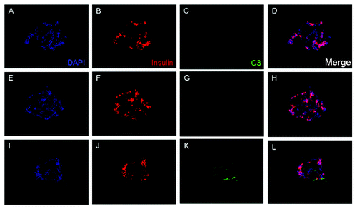
Figure 2. Complement activation initiated by auto-antibodies in type 1 diabetic serum induce a specific profile of gene expression. Islets exposed to complement in diabetic serum or control sera (allogenic or autologous) were compared using Illumina micro-array analysis. The resulting data was analyzed using the GeneSpring Software. Supervised clustering revealed 679 differentially expressed genes in diabetic serum when compared with autologous serum. Data from one representative experiment out of five independent experiments is shown.
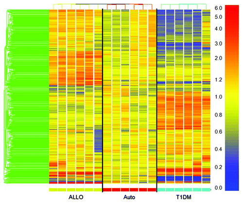
Figure 3. class prediction analysis of islets exposed to type 1 diabetic serum. One representative experiment was chosen for class prediction analysis to establish a list of genes that could differentiate the serum conditions based on gene expression changes. Samples were normalized to the mean of the autologous condition. Yellow reflects unchanged expression, blue reflects downregulation and red reflects upregulation.
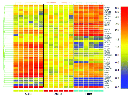
Table 1. Average signal intensity for class predictor genes in T1DM serum-treated islets
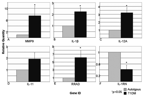
Figure 4. Real time quantitative PCR analysis confirms micro-array data in representative genes. Representative genes were chosen for corroboration by using relative quantification real-time PCR. TaqMan probes specific for MMP-9 (A), IL-1β (B), IL-12A (C), IL-11 (D), RAD (E), and IL-1RN (F) were used to produce cDNA from representative mRNA samples from each experiment. Real-time PCR detected significant gene expression changes of 7.50 ± 3.17 (p = 0.07), 2.23 ± 0.33 (p = 0.005), 3.19 ± 0.66 (p = 0.01), 1.92 ± 0.53 (p = 0.12), 6.13 ± 1.87 (p = 0.03), 0.42 ± 0.14 (p = 0.003) respectively. Data expressed as mean ± SEM.