Figures & data
Table 1. Animal characteristics.
Figure 1. The change in blood glucose concentration from pre to post exercise. The change in blood glucose concentrations in response to exercise was significantly different between non-T1DM exercised rats (CE) and T1DM exercised rats (DE; P < 0.05). A significant increase in blood glucose was evident in response to exercise in CE rats while a significant decrease was evident in DE rats (P < 0.05). The change in blood glucose concentration following exercise was not significantly changed at any point during training in CE or DE rats (P > 0.05). Data are expressed means ± SE for each animal group.
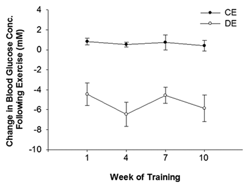
Figure 2. Representative images of pancreatic islet insulin staining (A) and insulin quantification (B). Pancreatic islets from both sedentary and exercised rats with T1DM (D and DE) had significantly less insulin staining than sedentary and exercised non-T1DM (C and CE) rats (P < 0.05). (*) indicates a significant difference from C. (#) indicates a significant difference from CE (P < 0.05). Data are expressed as means ± SE for each animal group. (x40 magnification; Bar = 50 µm)
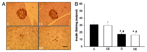
Figure 3. Representative images of pancreatic islet glucagon staining (A) and glucagon quantification (B). Pancreatic islets from both sedentary and exercised rats with T1DM (D and DE) had significantly more glucagon staining than sedentary and exercised non-T1DM (C and CE) rats (P < 0.05). (*) indicates a significant difference from C. (#) indicates a significant difference from CE (P < 0.05). Data are expressed as means ± SE for each animal group. (x40 magnification; Bar = 50 µm)
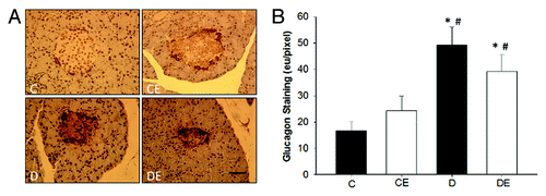
Figure 4. Pancreatic islet area. Pancreatic islets from both sedentary and exercised rats with T1DM (D and DE) were significantly smaller than sedentary and exercised non-T1DM (C and CE) rats (P < 0.05). (*) indicates a significant difference from C. (#) indicates a significant difference from CE (P < 0.05). Data are expressed as means ± SE for each animal group.
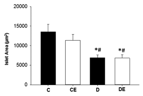
Figure 5. Pancreatic islet insulin (A) and glucagon (B) staining by diameter (small islets < 50 µm). Small islets had significantly more insulin and glucagon staining than larger islets. (*) indicates a significant difference (P < 0.05). Data are expressed as means ± SE for each animal group. (C: non-T1DM sedentary; CE; non-T1DM exercised; D: T1DM sedentary; DE: T1DM exercised)
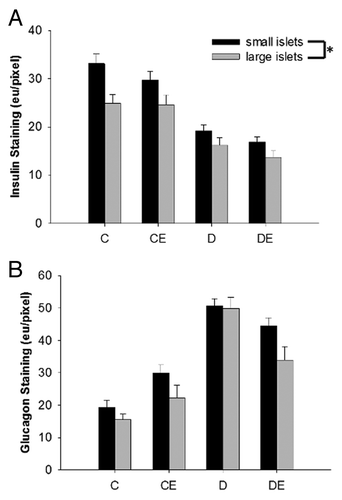
Figure 6. Representative Ki67 stain (A) and number of Ki67 positive cells per islet (B) based on islet diameter (small islets < 50 µm). Large islets had significantly more Ki67 positive cells (P < 0.05). (*) indicates a significant main effect of islet size. Data are expressed as means ± SE for each animal group. (C: non-T1DM sedentary; CE; non-T1DM exercised; D: T1DM sedentary; DE: T1DM exercised). (x40 magnification; Bar = 50µm)
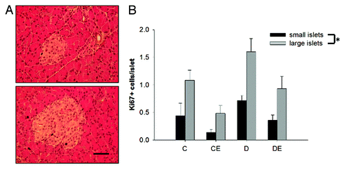
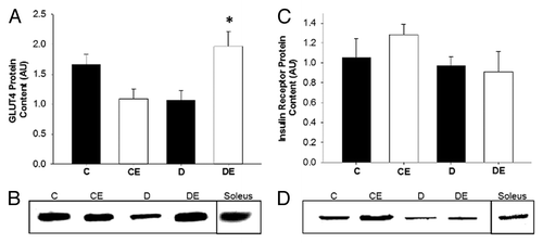
Figure 7. GLUT4 protein content in the red vastus muscle (A) and representative blot (B). Insulin receptor protein content in the red vastus muscle (C) and representative blot (D). Exercise training resulted in a significant increase in GLUT4 protein content in rats with T1DM. No change in insulin receptor protein content was evident across experimental groups. (*) indicates a significant difference compared with D. Data are expressed as means ± SE for each animal group. (C: non-T1DM sedentary; CE; non-T1DM exercised; D: T1DM sedentary; DE: T1DM exercised)