Figures & data
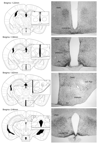
Figure 1. Images show leptin-induced phospho-STAT3 immunohistochemistry in coronal sections of the hypothalamus of fasted female mice treated with an intraperitoneal injection of leptin (1 mg/kg BW).Citation67 Hypothalamic regions displaying leptin-induced phospho-STAT3 include the ventromedial nucleus (VMN), the dorsal medial area of the VMN (VMNdm), arcuate nucleus (Arc), dorsomedial nucleus (DMN), lateral hypothalamus (Lat. Hyp) and ventral premammillary nucleus (PMV).