2,330
Views
34
CrossRef citations to date
0
Altmetric
Review

LIF-STAT signaling and trophoblast biology

, &
Article: e25155 | Received 02 May 2013, Accepted 23 May 2013, Published online: 27 Jun 2013

Figures & data

Figure 1. Significance of LIF-mediated signaling in blastocyst attachment. LIF is expressed by the receptive endometrial luminal and glandular epithelium. At the same time LIFR is expressed by the endometrial luminal epithelium as well as by the blastocyst. At the time of implantation, endometrial epithelial cells express integrin αVβ3 as well as osteopontin (not shown in figure) that forms the part of pinopodes essential for the initiation of implantation. A juxtacrine signaling through HB-EGF expressed on the endometrial epithelium leads to the expression of integrin α5β3 by trophoblast cells. These changes collectively bring out attachment of the blastocyst to the endometrium. Once the blastocyst gets attached, it also starts expressing LIF that can act in autocrine or paracrine way on trophoblast and endometrial cells, respectively.
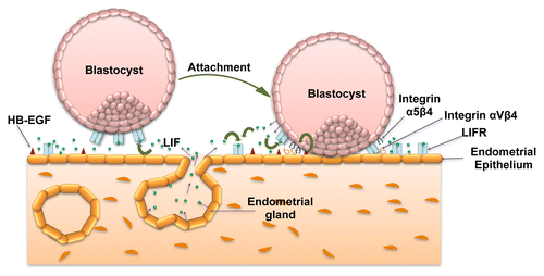
Figure 1. Significance of LIF-mediated signaling in blastocyst attachment. LIF is expressed by the receptive endometrial luminal and glandular epithelium. At the same time LIFR is expressed by the endometrial luminal epithelium as well as by the blastocyst. At the time of implantation, endometrial epithelial cells express integrin αVβ3 as well as osteopontin (not shown in figure) that forms the part of pinopodes essential for the initiation of implantation. A juxtacrine signaling through HB-EGF expressed on the endometrial epithelium leads to the expression of integrin α5β3 by trophoblast cells. These changes collectively bring out attachment of the blastocyst to the endometrium. Once the blastocyst gets attached, it also starts expressing LIF that can act in autocrine or paracrine way on trophoblast and endometrial cells, respectively.

Figure 2. STAT-dependent signaling and gene expression in LIF treated trophoblastic cells. LIF upon binding to the gp130-LIF receptor complex present on the plasma membrane of trophoblastic cells activate JAKs that ultimately phosphorylate the STAT3 and or STAT1 in the cytoplasm. These activated STATs form the homo- or hetero-dimers and move inside the nucleus to influence the expression of various genes that could regulate different functions like cytokine and signaling (IL-6, OSMR, SOCS3, and JUNB), adhesion (CECAM1, PDPN, and ITGB3), invasion (PAPPA, Caspase1, SERPINB3, TIMP1, TIMP2, and TIMP3), angiogenesis (ID1, ICAM1, EDIL3, and CCL2), etc. The genes whose expression is downregulated following LIF treatment are shown with down-arrow, while those showing upregulation are depicted with an up arrow.
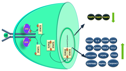
Figure 2. STAT-dependent signaling and gene expression in LIF treated trophoblastic cells. LIF upon binding to the gp130-LIF receptor complex present on the plasma membrane of trophoblastic cells activate JAKs that ultimately phosphorylate the STAT3 and or STAT1 in the cytoplasm. These activated STATs form the homo- or hetero-dimers and move inside the nucleus to influence the expression of various genes that could regulate different functions like cytokine and signaling (IL-6, OSMR, SOCS3, and JUNB), adhesion (CECAM1, PDPN, and ITGB3), invasion (PAPPA, Caspase1, SERPINB3, TIMP1, TIMP2, and TIMP3), angiogenesis (ID1, ICAM1, EDIL3, and CCL2), etc. The genes whose expression is downregulated following LIF treatment are shown with down-arrow, while those showing upregulation are depicted with an up arrow.