
Figures & data
Table 1. Characteristics of a 10 mg/ml solution of antibody in PBS pH 7.2 after nebulization with three nebulizers. Each result is the mean (± SD) of three nebulizations
Figure 1. Large antibody aggregates were analyzed by fluorescence microscopy before and after nebulization with the three nebulizers (Omron, PARI eFlow, Aerogen Solo), for a 10 mg/ml antibody solution.
Figure 2. A and 2B. Size exclusion chromatography of a 10 mg/ml antibody solution in PBS before (solid line) and after (dotted line) nebulization with the Aerogen Solo. (A) UV signal; (B) MALLS signal. The left is Molecular Weight determined by MALLS and the right axis is light scatter in arbitrary units.
Figure 3. The percentage increase in the number of large antibody aggregates was analyzed by fluorescence microscopy following nebulization, for the three nebulizers (Omron, PARI eFlow, Aerogen Solo).
Table 2. Minimum concentration of PS-20 required to prevent the production of medium-sized aggregates during the nebulization of an antibody solution (10 mg/ml in PBS pH 7.2) with three nebulizers
Table 3. Characteristics of a 10 mg/ml antibody solution in PBS pH 7.2 supplemented with various concentrations of PS-20, after nebulization with the Aerogen Solo nebulizer. Each result is the mean (± SD) of three nebulizations
Table 4. Characteristics of solutions of two monoclonal antibodies (IgG1 and IgG4) in different buffers with various concentrations of polysorbate, before and after nebulization. Each result is the mean (± SD) of three nebulizations
Table 5. Characteristics of solutions of various concentrations of antibody in PBS pH 7.2, after nebulization with the Aerogen Solo nebulizer. Each result is the mean (± SD) of three nebulizations
Figure 4. Large antibody aggregates were analyzed by fluorescence microscopy before and after nebulization with the Aerogen Solo nebulizer, for various antibody concentrations.
Figure 5. Model of the spatial distribution of antibodies in aerosol droplets as a function of antibody concentration.
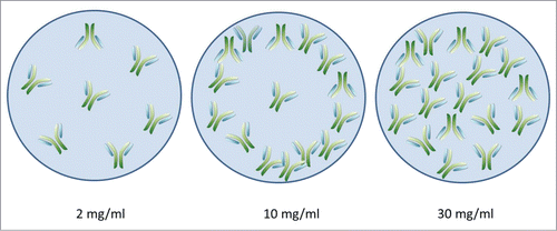
Table 6. Minimum concentration of PS-20 required to limit the formation of medium-sized and large aggregates