Figures & data
Figure 1. Supraependymal serotonergic axons are present in humans. (A and B) The lateral wall of the anterior horn of lateral ventricle (black arrows) in a 6-mo-old human brain was examined at the rostral (A) and caudal (B) levels. (C, F, and I) Immunostaining for acetylated tubulin (AcTub) reveals long axonal processes (white arrows) coursing among tufts of ependymal cilia. (C–H) These axons express both serotonin (5HT) (C–E) and serotonin transporter (SERT) (F–H). (I–K) Punctate expression of the presynaptic marker synaptophysin is observed at the varicosities (white arrows) along the axons.
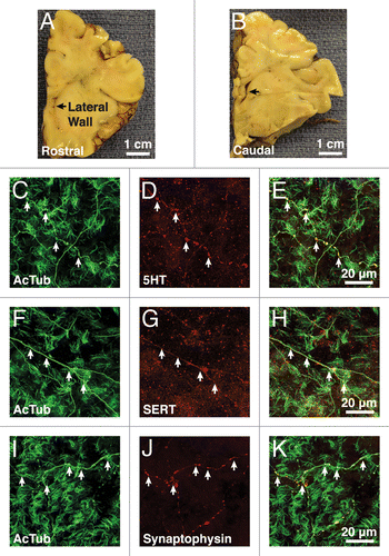
Figure 2. Supraependymal axons in humans have similar ultrastructures as the mice. (A) Tangential section of an axonal fiber shows long microtubules (black arrow) running parallel to the axonal shaft. The axon is closely associated with microvilli (white arrow) on the ependymal surface. (B) Transverse section of the axons reveals dark (black arrow) and light (white arrow)-core vesicles. (C) Supraependymal axonal varicosities also contain mitochondria (black arrows). (D) Note the invagination of the ependymal membrane (black arrow) next to the axons, suggestive of endo/exocytosis. (E and F) Symmetrical electron-dense contacts (black arrows) are observed between axons and ependymal (E1) cells. White arrow in (E) points to the base of an ependymal cilium.
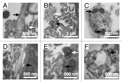
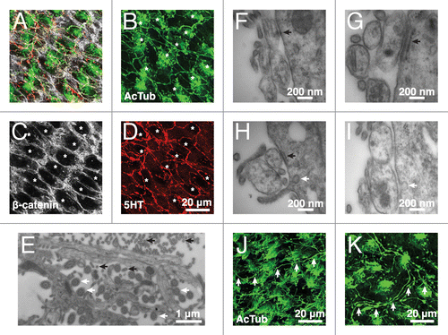
Figure 3. Supraependymal serotonergic axons make specialized contacts with E1 cells in mice. (A–D) Confocal microscopy shows supraependymal 5HT axons looping around the apical surfaces of E1 cells (indicated by asterisks). (E) EM of the V–SVZ wholemount shows that microvilli (black arrows) line the length of the axonal shafts. Ependymal cilia (white arrows) are identified by their characteristic (9+2) microtubule arrangement. (F) A transverse section shows a desmosome-like contact between a suprapendymal axon and a putative E1 cell. (G and H) A fragment of endoplasmic reticulum cisterna (black arrow) is frequently observed in the E1 cell adjacent to the site of contact with a supraependymal axon. (H and I) Membrane invaginations (white arrow) are also observed close to the site of contact between E1 cells and supraependymal axons, suggesting active endo/exocytosis. (J and K) Other than the lateral ventricles, antibodies against AcTub also stained supraependymal axons (white arrows) in the third (J) and fourth (K) ventricles of the mouse brain.