Figures & data
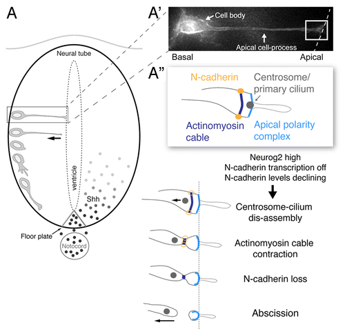
Figure 1. Steps underlying apical abscission in the neural tube. (A) Schematic of sequence of events taking place as a cell differentiates into a neuron in the early neural tube/spinal cord, beginning with the withdrawal of the apical cell-process from the ventricular surface and subsequent production of a growth cone and primary axon extension. Sonic hedgehog (Shh) signals from the notochord/floor plate promote proliferation and pattern in the spinal cord and their transduction requires an intact primary cilium; (A’) A cell poised to undergo neuronal differentiation, characterized by a basally located cell body and attachment to the apical surface through an elongated apical cell-process. (A”) Schematic of key sub-cellular structures in the apical endfoot of the prospective neuron (white boxed region in A’) (including apical complex containing apical membrane, the primary cilium, cadherin-containing adherens junctions and associated actinomyosin cable) and sequential changes leading to apical abscission following loss of N-cadherin.