Figures & data
Figure 1. Confocal images of endogenous UPF1 distribution in human HeLa cancer cells. Indirect immunofluorescence staining was performed using goat polyclonal antibodies raised against a C-terminal peptide of UPF1 on fixed cells either untreated, to detect total UPF1 (upper panels), or pre-treated with mild detergents, to detect insoluble UPF1 (lower panels). UPF1 antibodies were revealed using secondary antibodies conjugated with Alexa 488 fluorochrome (shown in green), whereas DNA was stained using DAPI (in blue). Note the focal staining of insoluble UPF1 in the nucleus. Scale bars correspond to 10 μm.
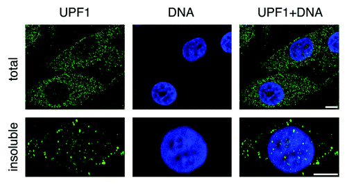
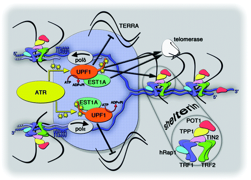
Figure 2. Speculative model depicting UPF1 association with a replicating telomere. Repetitive telomeric DNA, in blue, is composed of double stranded 5′-TTAGGG-3′/5′-CCCTAA-3′ repeats and it unwinds into single-stranded telomeric DNA within the replication fork, shown as a blue shaded ellipse. UPF1 physically interacts with the lagging-strand polymerase polδ and, possibly, with the leading-strand polymerase polε. Both polymerases are in gray and the arrows indicate the direction of replication on the two strands. The ATPase activity of UPF1 is indicated by the reaction ATP → ADP + inorganic phosphate (Pi). ATR (in yellow) phosphorylates UPF1, presumably within its C-terminal tail. Human EST1A (in green) directly interacts with UPF1. Black arrows indicate interactions between UPF1, hEST1A, TPP1 (in yellow within the shelterin complex), and telomerase (in white). The black inhibitory bar indicates the putative function associated with UPF1 in displacing TERRA from the replicating telomere.