1,122
Views
15
CrossRef citations to date
0
Altmetric
Research Paper

An inducible nuclear body in the Drosophila germinal vesicle

Pages 403-409 | Received 21 Jun 2011, Accepted 27 Jul 2011, Published online: 01 Sep 2011

Figures & data

Figure 1 Individual frames from a video of a stage-10 GV of Drosophila, showing gradual formation of induced nuclear bodies. The first frame (t = 0) was taken approximately five minutes after the egg chamber had been gently squashed between a glass slide and coverslip. In this GV the first bodies to form were numerous and irregular in outline, but eventually most coalesced into three large bodies. Arrow points to the karyosome, which remained unchanged. Imaged by DIC. Bar = 10 µm. A video of another GV can be watched in the Supplemental Material.

Figure 1 Individual frames from a video of a stage-10 GV of Drosophila, showing gradual formation of induced nuclear bodies. The first frame (t = 0) was taken approximately five minutes after the egg chamber had been gently squashed between a glass slide and coverslip. In this GV the first bodies to form were numerous and irregular in outline, but eventually most coalesced into three large bodies. Arrow points to the karyosome, which remained unchanged. Imaged by DIC. Bar = 10 µm. A video of another GV can be watched in the Supplemental Material.

Figure 2 Induced nuclear bodies in GVs from egg chambers of increasing size. In general, smaller GVs form only one or two bodies, whereas the largest GVs often display 15–20 bodies. Stage 7–8: a Cajal body (CB) is normally found in GVs up to about stage 8, at which time it disappears. The induced nuclear body (NB) in this stage 7–8 GV is independent of the CB, but is closely associated with the karyosome (K). Stage 9: nuclear bodies may contain vacuoles of trapped nucleoplasm, especially evident in this stage 9 GV. Stage 10: Multiple nuclear bodies are common in larger GVs. The karyosome (K) is in focus in this image. Stage 11: The needle-like inclusion (N) seen here occurs regularly in GVs and nurse cell nuclei of untreated oocytes. Bar = 10 µm.

Figure 2 Induced nuclear bodies in GVs from egg chambers of increasing size. In general, smaller GVs form only one or two bodies, whereas the largest GVs often display 15–20 bodies. Stage 7–8: a Cajal body (CB) is normally found in GVs up to about stage 8, at which time it disappears. The induced nuclear body (NB) in this stage 7–8 GV is independent of the CB, but is closely associated with the karyosome (K). Stage 9: nuclear bodies may contain vacuoles of trapped nucleoplasm, especially evident in this stage 9 GV. Stage 10: Multiple nuclear bodies are common in larger GVs. The karyosome (K) is in focus in this image. Stage 11: The needle-like inclusion (N) seen here occurs regularly in GVs and nurse cell nuclei of untreated oocytes. Bar = 10 µm.

Figure 3 Induced nuclear bodies in a GV from a transgenic line expressing EYFP-coilin. (A) DIC image. (B) Fluorescence image. The exclusion of EYFP-coilin from the bodies suggests that they are unrelated to CBs. Bar = 10 µm.
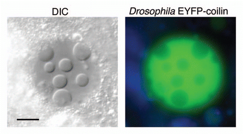
Figure 3 Induced nuclear bodies in a GV from a transgenic line expressing EYFP-coilin. (A) DIC image. (B) Fluorescence image. The exclusion of EYFP-coilin from the bodies suggests that they are unrelated to CBs. Bar = 10 µm.

Figure 4 (A) Electron micrograph of an induced nuclear body in a GV from a stage-10 oocyte. The internal vacuoles in the nuclear body appear to be pools of trapped nucleoplasm. Bar = 5 µm. (B) Higher magnification of part of another nucleus shows that the body contains irregular electron-dense particles with diameters in the range of 30–50 nm, suspended in material indistinguishable from the nucleoplasm. Note the clearly defined membranes of the nuclear envelope (NE) but the absence of a membrane surrounding the nuclear body. Bar = 0.5 µm.
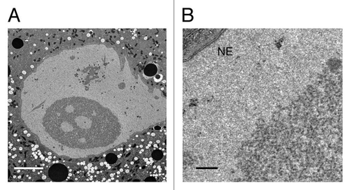
Figure 4 (A) Electron micrograph of an induced nuclear body in a GV from a stage-10 oocyte. The internal vacuoles in the nuclear body appear to be pools of trapped nucleoplasm. Bar = 5 µm. (B) Higher magnification of part of another nucleus shows that the body contains irregular electron-dense particles with diameters in the range of 30–50 nm, suspended in material indistinguishable from the nucleoplasm. Note the clearly defined membranes of the nuclear envelope (NE) but the absence of a membrane surrounding the nuclear body. Bar = 0.5 µm.

Figure 5 (A) GVs exposed to a solution containing only NaCl develop nuclear bodies. (B) Nuclear bodies do not form when GVs are exposed to Grace's insect culture medium. (C) Bodies fail to form when the isolation medium contains MgCl2 in the 10–30 mM range. The same is true for CaCl2. Bar = 10 µm.
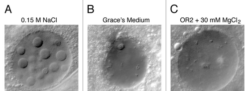
Figure 5 (A) GVs exposed to a solution containing only NaCl develop nuclear bodies. (B) Nuclear bodies do not form when GVs are exposed to Grace's insect culture medium. (C) Bodies fail to form when the isolation medium contains MgCl2 in the 10–30 mM range. The same is true for CaCl2. Bar = 10 µm.

Figure 6 Nuclear bodies were induced in lines from the Carnegie Protein Trap Library.Citation12 In each case, a specific protein is labeled with GFP, making it possible to determine whether that protein is included or excluded from the body. For protein abbreviations see the text and Flybase (http://flybase.org/). Twenty-two protein trap lines were examined, as shown in Table S1. Bar = 10 µm.
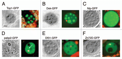
Figure 6 Nuclear bodies were induced in lines from the Carnegie Protein Trap Library.Citation12 In each case, a specific protein is labeled with GFP, making it possible to determine whether that protein is included or excluded from the body. For protein abbreviations see the text and Flybase (http://flybase.org/). Twenty-two protein trap lines were examined, as shown in Table S1. Bar = 10 µm.

Figure 7 FRAP analysis of GFP-labeled topoisomerase 1, a protein that is strongly localized in the induced nuclear bodies. (A) The first panel shows a GV that contains several large nuclear bodies. The largest of these (circle) was bleached at t = 0 s. Recovery from bleaching was rapid, as shown in images taken at 6.8 s, 101 s and 276 s. (B) Graph of the average intensity of the body throughout the experiment, normalized to a pre-bleach intensity of 1.0. The half time for recovery was 45 s and full recovery was reached at about 200 s. Bar = 10 µm.
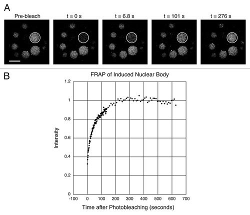
Figure 7 FRAP analysis of GFP-labeled topoisomerase 1, a protein that is strongly localized in the induced nuclear bodies. (A) The first panel shows a GV that contains several large nuclear bodies. The largest of these (circle) was bleached at t = 0 s. Recovery from bleaching was rapid, as shown in images taken at 6.8 s, 101 s and 276 s. (B) Graph of the average intensity of the body throughout the experiment, normalized to a pre-bleach intensity of 1.0. The half time for recovery was 45 s and full recovery was reached at about 200 s. Bar = 10 µm.

Figure 8 DIC image of a living mouse egg shortly before fusion of the two pronuclei. In this focal plane the female pronucleus displays a single large nuclear body, whereas the slightly larger male pronucleus shows one small and two large bodies. The polar body nucleus also appears to contain a small nuclear body. Note the superficial similarity to the induced nuclear bodies of Drosophila.
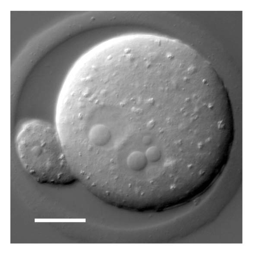
Figure 8 DIC image of a living mouse egg shortly before fusion of the two pronuclei. In this focal plane the female pronucleus displays a single large nuclear body, whereas the slightly larger male pronucleus shows one small and two large bodies. The polar body nucleus also appears to contain a small nuclear body. Note the superficial similarity to the induced nuclear bodies of Drosophila.