410
Views
0
CrossRef citations to date
0
Altmetric
Extra View

Defining flexible vs. inherent promoter architectures

The importance of dynamics and environmental considerations

&
Pages 399-403 | Published online: 03 Jul 2012

Figures & data

Figure 1. Promoter architecture is influenced by NDR placement. Nucleosome occupancy 400 bp upstream to the TSS, in unstressed cells (left) or 60 min after treatment with 0.4 mM H2O2 (right) from.Citation8 Upstream nucleosome occupancy is shown for 493 DPN genes (A) or 543 OPN genes (B), as defined by Tirosh and Barkai;Citation14 genes were organized by hierarchical clustering of the upstream regions. Occupancy is shown on the same scale for both gene groups, according to the key.
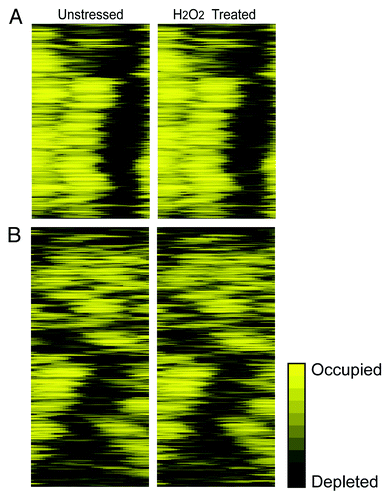
Figure 1. Promoter architecture is influenced by NDR placement. Nucleosome occupancy 400 bp upstream to the TSS, in unstressed cells (left) or 60 min after treatment with 0.4 mM H2O2 (right) from.Citation8 Upstream nucleosome occupancy is shown for 493 DPN genes (A) or 543 OPN genes (B), as defined by Tirosh and Barkai;Citation14 genes were organized by hierarchical clustering of the upstream regions. Occupancy is shown on the same scale for both gene groups, according to the key.

Figure 2. Two classes of promoters based on NDR accessibility. Data are shown as described in . (A) 1,069 genes with no nucleosome signal in unstressed cells, before (left) and 60 min after treatment with 0.4 mM H2O2 (right) from.Citation8 (B) 288 genes whose promoters rank in the top 5% based on nucleosome signal in the NDR. Genes in both classes were organized by position of the NDR. The contrast was increased for genes in (B) to distinguish the NDR.
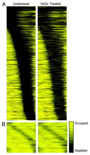
Figure 2. Two classes of promoters based on NDR accessibility. Data are shown as described in Figure 1. (A) 1,069 genes with no nucleosome signal in unstressed cells, before (left) and 60 min after treatment with 0.4 mM H2O2 (right) from.Citation8 (B) 288 genes whose promoters rank in the top 5% based on nucleosome signal in the NDR. Genes in both classes were organized by position of the NDR. The contrast was increased for genes in (B) to distinguish the NDR.