Figures & data
Figure 1. Intracellular localizations and dynamics of progerin and non-farnesylated progerin in transiently transfected MEFs. (A) Confocal fluorescence micrographs showing localizations of GFP-progerin at the nuclear envelope and GFP-progerin-NF in intranuclear foci. The arrow indicates the irregular shape, and the arrowhead points a nuclear envelope bleb in the cells expressing GFP-progerin. Bar: 5 µm. (B) The diffusional mobility of GFP-progerin-NF is higher than that of GFP-progerin in transfected MEFs. Quantitative experiments showing normalized fluorescence recovery after photobleaching in cells transiently transfected with cDNA constructs encoding GFP-progerin (red) or GFP-progerin-NF (green). Normalized fluorescence of 1 is the level before bleaching. Normalized fluorescence for GFP-progerin-NF is statistically significantly higher than that for GFP-progerin (p < 0.05 at 13 sec, 22 sec, 31 sec, 61 sec, 102 sec, 302 sec and 654 sec these time points; n = 12 cells analyzed for GFP-progerin and n = 10 cells analyzed for GFP-progerin-NF). Values shown are means plus or minus standard deviations. (C) Confocal immunofluorescence micrographs of transiently transfected MEFs expressing GFP-progerin-NF (green) and labeled with antibodies against RNAPII, PML or PCNA (red) with signal overlap appearing yellow (merge). Bar: 5 µm.
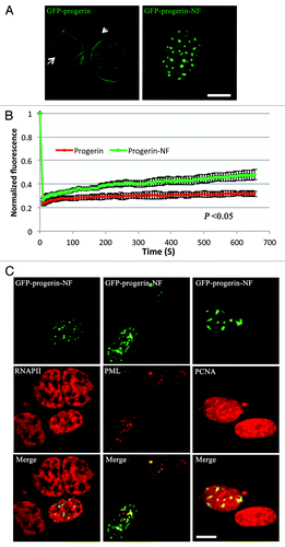
Figure 2. Expression of non-farnesylated progerin alters the nuclear distribution of lamin A in transiently transfected MEFs. Confocal fluorescence micrographs showing localizations of GFP-progerin and GFP-progerin-NF (green signals) and immunofluorescence labeling with anti-lamin A antibodies (red signals) in the same cells; merged images are shown at the right with signal overlap appearing yellow (merge). Bar: 5 µm.
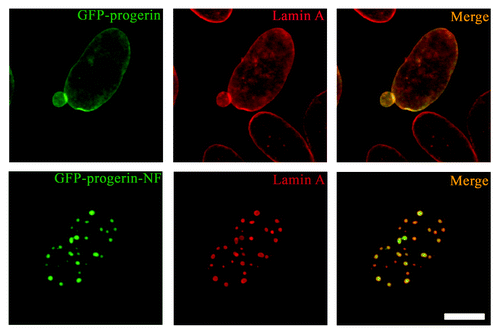
Figure 3. Expression of non-farnesylated progerin alters the nuclear distribution of lamin C in transiently transfected MEFs. Confocal fluorescence micrographs showing localizations of GFP-progerin and GFP-progerin-NF (green signals) with RFP-lamin C (red signals) in the same cells; merged images are shown at the right with signal overlap appearing yellow (merge). Bar: 5 µm.
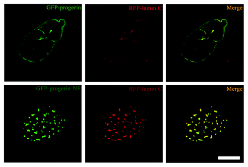
Figure 4. Expression of non-farnesylated progerin does not significantly alter the nuclear distribution of lamin B1 in transiently transfected MEFs. Confocal fluorescence micrographs showing localizations of GFP-progerin and GFP-progerin-NF (green signals) and immunofluorescence labeling with anti-lamin B1 antibodies (red signals) in the same cells; merged images are shown at the right with signal overlap appearing yellow (merge). Bar: 5 µm.
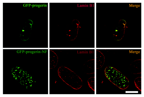
Figure 5. Effects of FTI treatment on protein farnesylation, progerin localization and intranuclear distribution of lamins. (A) FTI treatment blocks protein farnesylation in MEFs. MEFs were incubated for 48 h with 0 µM or 10 µM FTI-276 and extracted proteins were separated by SDS-PAGE and immunoblotted with antibodies against lamin A/C, HDJ-2 and β-actin. (B) Treatment of MEFs with FTI leads to intranuclear localization of GFP-progerin and altered distributions of lamin A and lamin C but not lamin B1. Confocal fluorescence micrographs showing localizations of GFP-progerin (green signals), immunofluorescence labeling with anti-lamin A antibodies (red signals, top row), immunofluorescence labeling with anti-lamin B1 antibodies (red signals, middle row) and RFP-lamin C (red signals, bottom row) in cells that were incubated with 10 µM FTI-276; merged images are shown at the right with signal overlap appearing yellow (merge). Bar: 5 µm.
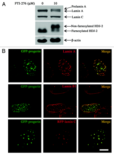
Figure 6. Effects of expressing non-farnesylated progerin on the distribution of two integral proteins of the inner nuclear membrane and non-membrane nuclear pore complex (NPC) proteins. (A) Confocal fluorescence micrographs showing localizations of GFP-progerin and GFP-progerin-NF (green signals) and immunofluorescence labeling with anti-LAP1 antibodies (red signal). (B) Confocal fluorescence micrographs showing localizations of GFP-progerin and GFP-progerin-NF (green signals) and immunofluorescence labeling with anti-emerin antibodies (red signals). (C) Confocal fluorescence micrographs showing localizations of GFP-progerin and GFP-progerin-NF (green signals) and immunofluorescence labeling with antibodies that recognize NPC proteins (red signals). Merged images of the same cells are shown at the right of each panel with signal overlap appearing yellow (merge). Bar: 5 µm.
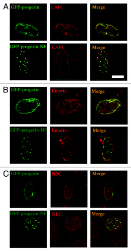
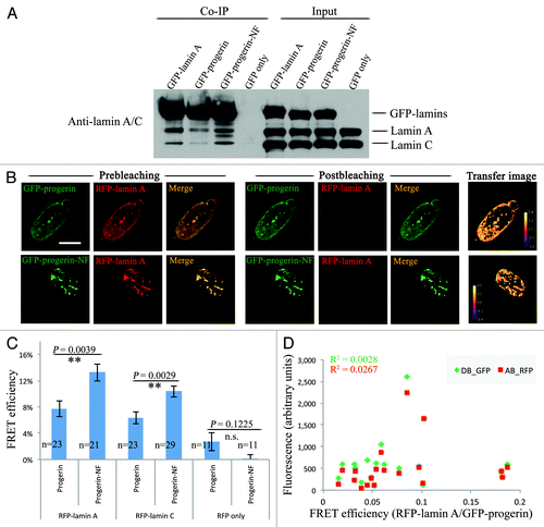
Figure 7. Binding of non-farnesylated progerin to A-type lamins. (A) Immunoblot with anti-lamin A/C antibodies of co-immunoprecipitated proteins (Co-IP) and inputs (Input) of cell extracts. MEFs transiently expressing GFP-lamin A, GFP-progerin, GFP-progerin-NF or GFP only were subjected to immunoprecipitation with antibody against GFP. Less endogenous lamin A and lamin C was precipitated from MEFs expressing GFP-progerin than GFP-lamin A or GFP-progerin-NF and no endogenous lamin A or lamin C was detected in precipitates from MEFs expressing only GFP. (B) Top row shows fluorescence micrographs of a cell expressing GFP-progerin (green) and RFP-lamin A (red) with merged image (yellow) and bottom row shows a cell expressing GFP-progerin-NF (green) and RFP-lamin A (red) with merged image (yellow). Images before photobleaching (Prebleaching), after photobleaching (Postbleaching) and energy transfer image (Transfer image) are indicated. Bar: 5μm. (C) Energy transfer efficiency between GFP progerin or GFP-progerin-NF and RFP-lamin A, RFP-lamin C or RFP only. Transfer efficiencies were significantly higher for GFP-progerin NF than for GFP-progerin (values shown are means plus or minus standard errors; p values calculated using Student’s t-test, two-tailed, two-sample unequal variance). No significant difference (n.s.) was detected in energy transfer efficiency between RFP only and GFP-progerin or GFP-progerin-NF. (D) Correlation of energy transfer efficiency and corresponding RFP-lamin A (acceptor) or GFP-progerin (donor) fluorescence intensity before photobleaching. AB_RFP: acceptor before photobleaching (red squares); DB-GFP: donor before photobleaching (green diamonds). No correlation was observed between FRET efficiencies and GFP-progerin or RFP-lamin A fluorescence intensity before photobleaching; similar lack of correlation was seen between FRET efficiencies and GFP-progerin-NF or RFP-lamin C fluorescence intensity before photobleaching (not shown).