Figures & data
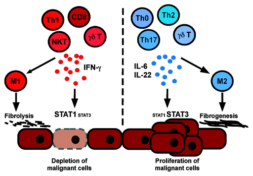
Figure 1. A model of cancer-promoting and cancer-inhibitory liver inflammation. Tumor-suppressive inflammatory liver infiltrates are characterized by high content of IFNγ-secreting lymphocytes (Th1, CD8 T, NK and NKT cells), sustained activation of the STAT1 pathway in hepatocytes, macrophage polarization toward an M1 phenotype and fibrolysis. In contrast, tumor-promoting liver infiltrates are characterized by high content of IFNγ non-producing or interleukin-22 producing lymphocytes, interleukin-6 secretion by various cell types, macrophages with M2 phenotype, sustained STAT3 activation in hepatocytes, fibrogenesis and angiogenesis.