Figures & data
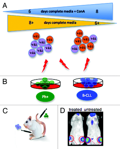
Figure 1. (A) Gamma delta T cells (GDTc) isolated from human blood were cultured for six to eight days in complete medium containing Concanavalin A (ConA). Vdelta1 (Vδ1) or Vdelta2 (Vδ2) subset prevalence in expanded culture correlated with the duration of exposure to ConA. (B) Vδ2 GDTc are cytotoxic to EM-2eGFPluc cells, Ph+ leukemia cells transduced with a lentiviral vector encoding eGFP and luciferase. Both Vδ1 and Vδ2 are cytotoxic to B-CLL-derived MEC1 and TMD2 cell lines. (C) The bioluminescent xenograft Ph+ leukemia model was established via intravenous (IV) injection of EM-2eGFPluc cells. Vδ2 GDTc therapy was investigated; infusion of GDTc intraperitoneally gave rise to better engraftment than when GDTc were injected IV. (D) Leukemia progression was monitored in vivo via IVIS® technology (In Vivo Imaging System, Xenogen). Shown here are representative examples of treated and leukemia-bearing mice. Red circles are regions of interest from which bioluminescence values were quantified.