Figures & data
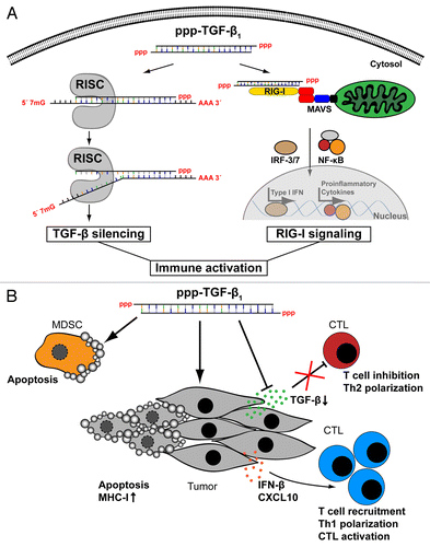
Figure 1. Immune activation with a triphosphate small-interfering RNA targeting transforming growth factor β brakes tumor-induced immunosuppression. (A) A 5′-triphosphate-modified small-interfering RNA targeting transforming growth factor β (ppp-TGFβ) combines the potential of RNA-interference (RNAi)-mediated TGFβ silencing and that of retinoic acid-inducible gene I (RIG-I) activation, leading to the secretion of type I interferon (IFN) and other pro-inflammatory cytokines. (B) In vivo, the administration of ppp-TGFβ leads to the production of type I IFN and various chemokines (such as CXCL10), to the upregulation of MHC class I expression on tumor cells as well as to tumor cell apoptosis. Additionally, ppp-TGFβ favors the apoptotic demise of myeloid-derived suppressor cells (MDSCs), the recruitment of CD8+ T cells into neoplastic lesions, TH1 polarization and cytotoxic T lymphocyte (CTL) activation in a murine model of pancreatic carcinoma.