Figures & data
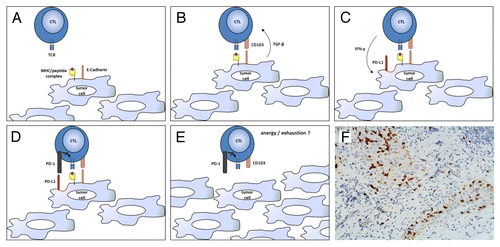
Figure 1. CD103+ lymphocytes infiltrating human ovarian carcinoma. (A–E) Ovarian cancer-infiltrating lymphocytes (TILs) express CD103 in response to tumor-associated antigen (TAAs) and transforming growth factor-β (TGF-β). CD103+ tumor-infiltrating lymphocytes (TILs) efficiently control tumor growth for a while, but then become trapped within neoplastic lesions as a consequence of CD103 expression, eventually becoming exhausted (PD-1+) owing to chronic antigen stimulation. (F) CD103+ TILs in a high-grade serous ovarian cancer specimen obtained from cytoreductive surgery. The section was stained with an anti-CD103 rabbit monoclonal antibody (Epitomics clone EPR4166Citation2), an anti-rabbit horseradish peroxidase-conjugate antibody and diaminobenzidine (DAB). CD103+ TILs (stained in brown) can be seen clustering within epithelial tumor regions. CTL, cytotoxic T lymphocyte; IFNγ, interferon γ; PD-L1, PD-1 ligand 1; TCR, T-cell receptor.