Figures & data
Figure 1. Schematic diagram of tissue engineering from amniotic fluid-derived stem cells for the treatment of congenital anomalies. Autologous fetal stem cells are obtained by amniocentesis. The cells are expanded ex vivo in parallel with the remainder of gestation and subsequently placed on biodegradable scaffolds prior to implantation at birth. Reprinted with permission.Citation5
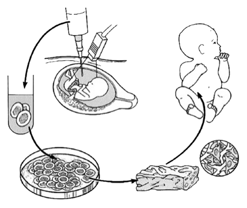
Figure 2. Gross appearance of a tissue-engineered heart valve seeded with amniotic fluid-derived stem cells. Reprinted with permission.Citation42
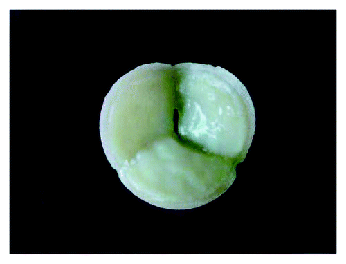
Figure 3. Gross appearance of a tissue-engineered diaphragmatic patch composed of amniotic fluid cells resuspended in a collagen hydrogel on acellular human dermis. Courtesy of Dario O. Fauza, Boston, MA USA.
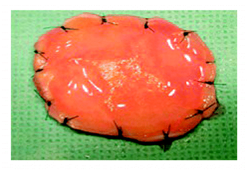
Figure 4. Three-dimensional micro-CT scan of a repaired full thickness sternal defect (contained within the black oval) using an amniotic fluid mesenchymal stem cell-based osseous construct. Reprinted with permission.Citation49
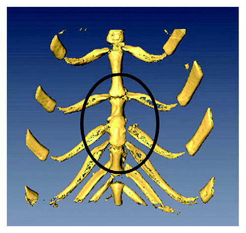
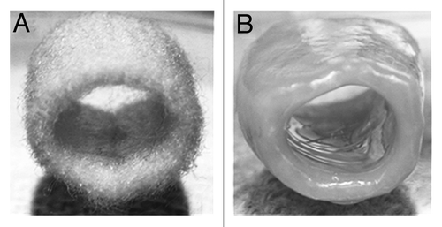
Figure 5. Gross appearance of a three-dimensional polyglycolic acid-based tubular scaffold before (A) and after (B) seeding with amniotic fluid-derived mesenchymal stem cells under chondrogenic conditions after 15 weeks in culture. Reprinted with permission.Citation51