Figures & data
Table 1: Definition of terms and common examples
Figure 1: (A) AP Radiograph of a hypertrophic nonunion of a subtrochanteric femur fracture, eleven months following cephalomedullary rod fixation. Coronal (B) and sagittal (C) CT images of the hypertrophic nonunion. (D) AP radiograph of the knee, showing insufficient effectively-unicortical screw fixation, which led to excess implant motion at the fracture site.

Figure 2: (B) PA and (C) lateral radiographs of a distal humeral atrophic nonunion, thirteen-months following open reduction and internal fixation of a comminuted, open distal humerus fracture. (A) Extensive soft tissue damage and periosteal stripping at the time of injury likely contributed to inadequate vasculature and for healing.
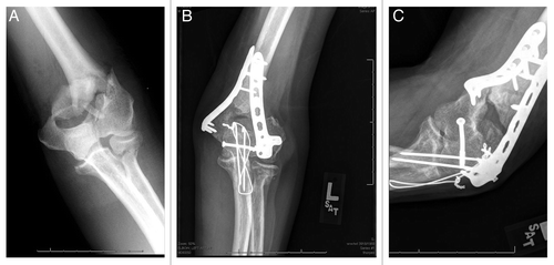
Table 2: Properties, functions and costs of various forms of bone grafts and substitutes
Figure 3: AP radiograph of a periprosthetic femur fracture following cable fixation augmented with a cortical strut femoral allograft.
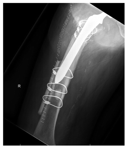
Table 3: Bone substitutes: properties and commercial product examples
Table 4: Select endogenous growth factors and their known functions in fracture healing
Table 5: Select Bone morphogenetic proteins: properties, indications and product examples
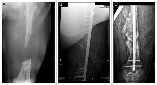
Figure 4: (A) AP radiograph of a large, segmental femur fracture with a sizeable diaphyseal defect. (B) postoperative AP radiograph of the fracture following bridge plating and implantation of iliac crest bone graph, DBM and BMP. (C) AP radiograph taken ten months following injury showing expansive bone formation and subsequent hypertrophic nonunion following hardware failure, treated with rigid intramedullary fixation.