Figures & data
Figure 1. Morphology of porous collagen scaffolds crosslinked with heparin (Col-Hep) (scanning electron microscopical images). Air-side and pan-side represent the side exposed to the air or to the mold while producing the scaffold, respectively. Bar represents 200 µm.
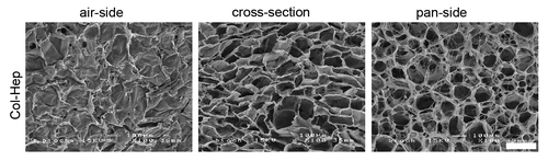
Figure 2. Immunolocalization of heparin and growth factors, and quantification of growth factors. (A) Heparin and growth factors were primarily located at the outer sites of the scaffolds, as evidenced by toluidin blue (heparin) and immunofluorescent staining (VEGF, HGF) of a Col-Hep scaffold and a Col-Hep-VH scaffold. Bars represent 200 µm. (B) Western blot for VEGF bound to Col-Hep-V and Col-Hep-VH scaffolds, and table indicating the amount of growth factors bound to scaffolds. Values depicted as mean of three experiments ± standard deviation. N/A, not applicable; Hep, heparin; V, VEGF; H, HGF.
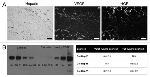
Figure 3. Macroscopical view of Col-Hep (A) and Col-Hep-H (B) scaffolds at 12 weeks after implantation. Arrows point at blood vessels at the lung side of Col-Hep-H scaffolds, which were not observed on Col-Hep scaffolds. Blue lines are non-absorbable sutures. Hep he,parin; H, HGF.
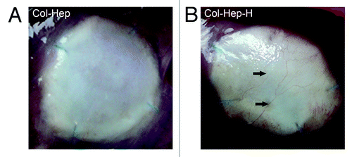
Table 1. Cells present in scaffolds at two and 12 weeks
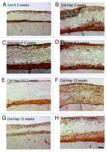
Figure 4. Vascularization of scaffolds (laminin staining). At two weeks, the scaffold substituted with heparin (Col-Hep; B) contained more blood vessels than other scaffolds (A, C–E). No significant differences between scaffolds types were observed at 12 weeks after implantation (F–H), although some variation between scaffolds was visible in distribution of blood vessels, both between and within groups. Bar represents 200 µm. Hep, heparin; V, VEGF; H, HGF.