Figures & data
Figure 1. Schema of our strategy of small intestinal PCS with cell transplantation. The PCS is constructed using an autologous small intestinal segment. Isolated hepatocytes are transplanted into the small intestinal graft.
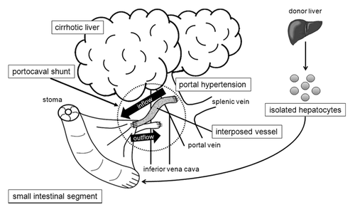
Figure 2. Luminescence from transplanted hepatocytes in rat small intestinal segments. Luminescence was stably detected for 30 d after transplantation.
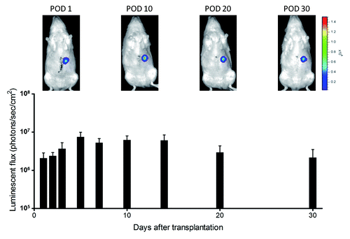
Figure 3. Histological analyses of intestinal segments in pigs. Intestinal segments were sampled on postoperative day 7. (A) Hepatocyte cluster in the submucosa (HE, x400). (B) Bile duct-like structure in the submucosa (HE, x400). (C) Glycogen in the hepatocytes (PAS, x200). (D) Immunohistochemical staining for albumin (x400). (E) Immunohistochemical staining for CYP1A1 (x400). (F) Immunohistochemical staining for OV-6 (x400).
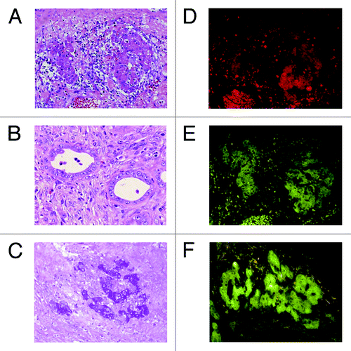
Figure 4. Intraoperative and postoperative findings of intestinal PCS in pigs. (A) Intraoperative image of the ileal graft and interposed vessel graft anastomosed to the host PV. (B) Intraoperative portography shows that temporary PH created by clamping the PV led to the visualization of the mesenteric artery of the intestinal graft. (C) The graft vessel interposed between the portal vein and small intestinal segment is occupied with a thrombus. (D) HE staining shows shortened villi and a thinned mucosal layer of the small intestinal segment.
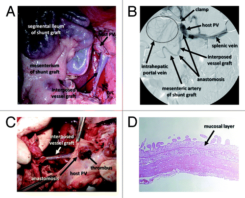
Table 1. Operative procedure and findings of the PCS experiment in pigs
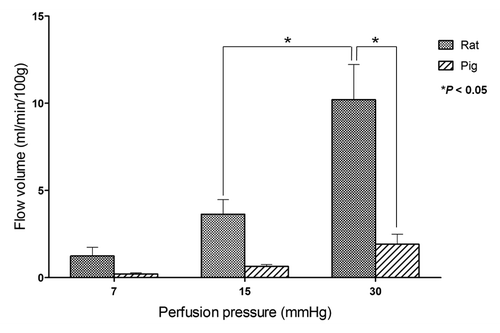
Figure 5. Relationship between flow volume and perfusion pressure in whole intestine ex vivo in rats and pigs. The mean flow volumes of the rat small intestine were larger than those of the pig under each pressure. *P value < 0.05.