3,581
Views
96
CrossRef citations to date
0
Altmetric
Review

The role of primary cilia in the development and disease of the retina

, &
Pages 69-85 | Received 10 Jun 2013, Accepted 04 Oct 2013, Published online: 25 Oct 2013

Figures & data

Figure 1. Schematic representation of a rod photoreceptor cell and localization of ciliary proteins.(A) The schematic represents the rod photoreceptor cell outer segment, connecting cilium, inner segment, outer fiber, cell body, inner fiber and synaptic terminus. A number of key components of the ciliary apparatus are color coded and indicated. The IFT complex A (blue) and complex B (red) are represented in the magnified inset. A retinal pigmentary epithelial (RPE) cell is shown in gray at the top. (B) Confocal microscopy images of an immunofluorescent stained P20 mouse retinal cryosection showing the stratified layers of the retina. Cilium transition zone and basal body protein MKS1 is stained in green, and a novel interactant of MKS1, RNF34, is stained in red. These proteins localize to the base of the connecting cilium, as shown by the arrowheads in the enlarged insets. (C) Confocal microscopy image of a human adult retinal pigment epithelium (ARPE19) cell overexpressing enhanced-GFP-tagged lebercilin and immunostained with an antibody against acetylated α tubulin, which marks the axoneme of the cilium. Lebercilin can be seen in a punctuate pattern along the axoneme. Scale bar = 10μm.
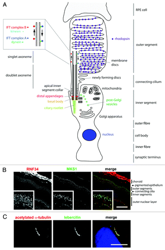
Figure 1. Schematic representation of a rod photoreceptor cell and localization of ciliary proteins.(A) The schematic represents the rod photoreceptor cell outer segment, connecting cilium, inner segment, outer fiber, cell body, inner fiber and synaptic terminus. A number of key components of the ciliary apparatus are color coded and indicated. The IFT complex A (blue) and complex B (red) are represented in the magnified inset. A retinal pigmentary epithelial (RPE) cell is shown in gray at the top. (B) Confocal microscopy images of an immunofluorescent stained P20 mouse retinal cryosection showing the stratified layers of the retina. Cilium transition zone and basal body protein MKS1 is stained in green, and a novel interactant of MKS1, RNF34, is stained in red. These proteins localize to the base of the connecting cilium, as shown by the arrowheads in the enlarged insets. (C) Confocal microscopy image of a human adult retinal pigment epithelium (ARPE19) cell overexpressing enhanced-GFP-tagged lebercilin and immunostained with an antibody against acetylated α tubulin, which marks the axoneme of the cilium. Lebercilin can be seen in a punctuate pattern along the axoneme. Scale bar = 10μm.

Table 1. The most comprehensively characterized photoreceptor proteins mutated in human disease

Supplemental material

Additional material

Download Zip (47.3 KB)

Reprints and Corporate Permissions

Please note: Selecting permissions does not provide access to the full text of the article, please see our help page How do I view content?

To request a reprint or corporate permissions for this article, please click on the relevant link below:

Academic Permissions

Please note: Selecting permissions does not provide access to the full text of the article, please see our help page How do I view content?

Obtain permissions instantly via Rightslink by clicking on the button below:

If you are unable to obtain permissions via Rightslink, please complete and submit this Permissions form. For more information, please visit our Permissions help page.