Figures & data
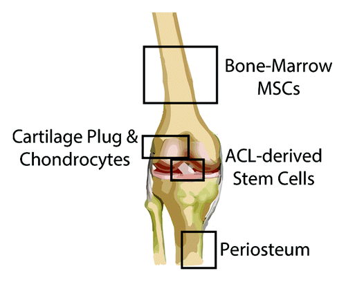
Figure 1. Histological features of the fibrocartilaginous bone-tendon interface stained with (A) Safranin-O, (B) H&E, and viewed with (C) polarized light microscopy. Note the four zones of the native enthesis. Magnification 20×. B, bone; CFC, calcified fibrocartilage; UFC, uncalcified fibrocartilage; T, tendon. Reproduced with permission from reference Citation122.
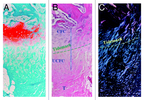
Figure 2. Embryonic development of the bone-tendon interface. (A) The degree of Scx and Sox9 expression determines cellular phenotype. Scx−/Sox9+ chondroprogenitors (CP) become chondrocytes of the skeletal anlagen while Scx+/Sox9− tenoprogenitors (TP) become tenocytes of the tendon midsubstance. Varying levels of Scx and Sox9 expression are seen in teno-/ligamento-/chondro-progenitors (TLCP), which give rise to cells of the bone-tendon interface. (B) Scx+/Sox9+ progenitors give rise to the primordial chondro-tendinous/ligamentous junction (CTJ/CLJ), which forms the osteo-tendinous/ligamentous junction (OTJ/OLJ) following birth. Reproduced with permission from reference Citation23.
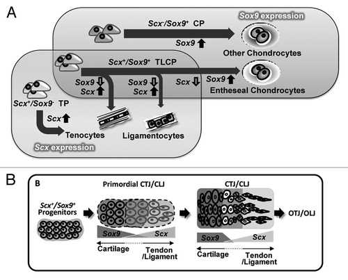
Figure 3. Cell sources for augmenting bone-tendon healing. Animal studies exploring the use of MSCs to improve bone-tendon healing have derived these cells primarily from the bone marrow, although other tissues (e.g., adipose) also harbor MSCs (not shown). ACL-derived MSCs can be obtained from the ruptured ligament, while chondrocytes/cartilage plugs can be taken from non-weightbearing articular surfaces. Lastly, the periosteum is typically harvested from the anteromedial tibia, given the ease of access in the absence of overlying soft tissues.