808
Views
15
CrossRef citations to date
0
Altmetric
Research Paper

Time-dependent expression of cytochrome p450 epoxygenases during human prenatal development

, , &
Pages 53-61 | Received 28 Aug 2013, Accepted 19 Jan 2014, Published online: 03 Feb 2014

Figures & data

Table 1. Brief summary of intestine, liver and kidney IUD events

Figure 1. Expression of CYP2C8, CYP29, CYP2C19, and CYP2J2 in enterocytes of embryonic/fetal intestine during 7th-20th week of IUD (n = 29) and adult duodenal tissue samples (n = 5). Graphs show the average histoscore. The error bars represent standard deviation.
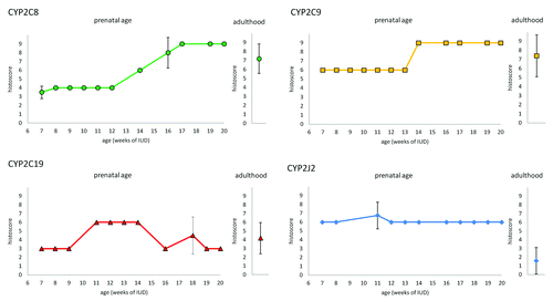
Figure 1. Expression of CYP2C8, CYP29, CYP2C19, and CYP2J2 in enterocytes of embryonic/fetal intestine during 7th-20th week of IUD (n = 29) and adult duodenal tissue samples (n = 5). Graphs show the average histoscore. The error bars represent standard deviation.

Figure 2. Differences between signal localization in fetal intestine in 16th week of IUD. CYP2C8 (A) and CYP2C9 (B) in enterocytes show stronger positivity in crypts than in apex of villi. On the other hand, CYP2C19 (C) and CYP2J2 (D) show stronger positivity in apex of villi, area of crypts is almost negative.
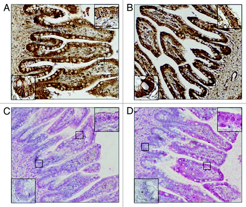
Figure 2. Differences between signal localization in fetal intestine in 16th week of IUD. CYP2C8 (A) and CYP2C9 (B) in enterocytes show stronger positivity in crypts than in apex of villi. On the other hand, CYP2C19 (C) and CYP2J2 (D) show stronger positivity in apex of villi, area of crypts is almost negative.

Figure 3. Expression of CYP2C8, CYP29, CYP2C19, and CYP2J2 in hepatocytes of embryonic/fetal liver during 5th-20th week of IUD (n = 23) and adult tissue samples (n = 5). Graphs show the average histoscore. The error bars represent standard deviation.
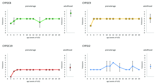
Figure 3. Expression of CYP2C8, CYP29, CYP2C19, and CYP2J2 in hepatocytes of embryonic/fetal liver during 5th-20th week of IUD (n = 23) and adult tissue samples (n = 5). Graphs show the average histoscore. The error bars represent standard deviation.

Figure 4. Embryonic kidney in 7th week of IUD. The proximal tubuli shows stronger staining than other type of tubuli. The metanephrogenic blastema shows weak positivity whereas uninduced mesenchyme is negative.
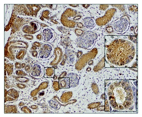
Figure 4. Embryonic kidney in 7th week of IUD. The proximal tubuli shows stronger staining than other type of tubuli. The metanephrogenic blastema shows weak positivity whereas uninduced mesenchyme is negative.

Figure 5. Expression of CYP2C8, CYP29, CYP2C19, and CYP2J2 in the proximal tubuli of embryonic/fetal kidney during 6th-20th week of IUD (n = 25) and adult tissue samples (n = 5). Graphs show the average histoscore. The error bars represent standard deviation.
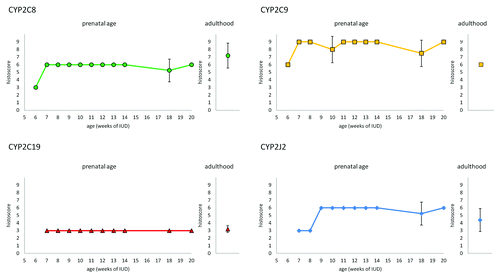
Figure 5. Expression of CYP2C8, CYP29, CYP2C19, and CYP2J2 in the proximal tubuli of embryonic/fetal kidney during 6th-20th week of IUD (n = 25) and adult tissue samples (n = 5). Graphs show the average histoscore. The error bars represent standard deviation.
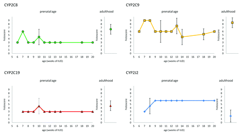
Figure 6. Expression of CYP2C8, CYP29, CYP2C19, and CYP2J2 in other tubuli (loops of Henle, distal tubuli, and collecting ducts) of embryonic/fetal kidney during 6th-20thweek of IUD (n = 25) and adult tissue samples (n = 5). Graphs show the average histoscore. The error bars represent standard deviation.

Figure 6. Expression of CYP2C8, CYP29, CYP2C19, and CYP2J2 in other tubuli (loops of Henle, distal tubuli, and collecting ducts) of embryonic/fetal kidney during 6th-20thweek of IUD (n = 25) and adult tissue samples (n = 5). Graphs show the average histoscore. The error bars represent standard deviation.

Table 2. Number of embryonic/fetal tissue samples in different ages of intrauterine development

Table 3. Number of used adult tissue samples and the basic characterization of donors (age, gender)

Supplemental material

Additional material

Download Zip (106 KB)

Reprints and Corporate Permissions

Please note: Selecting permissions does not provide access to the full text of the article, please see our help page How do I view content?

To request a reprint or corporate permissions for this article, please click on the relevant link below:

Academic Permissions

Please note: Selecting permissions does not provide access to the full text of the article, please see our help page How do I view content?

Obtain permissions instantly via Rightslink by clicking on the button below:

If you are unable to obtain permissions via Rightslink, please complete and submit this Permissions form. For more information, please visit our Permissions help page.