1,587
Views
15
CrossRef citations to date
0
Altmetric
Research Paper

Loss of cilia causes embryonic lung hypoplasia, liver fibrosis, and cholestasis in the talpid3 ciliopathy mutant

, , , &
Pages 177-185 | Received 04 Jan 2014, Accepted 08 Apr 2014, Published online: 17 Apr 2014

Figures & data

Figure 1. The talpid3 chicken exhibits abnormal lung and liver morphology. Compared with E10 wt lung (A), the talpid3 lung is smaller and poorly branched (B), air sac development was normal (red asterisk) (A andB). Wt liver (C) and talpid3 liver (D) are of similar size, although the talpid3 liver is green. The wt gall bladder is bile filled (green) (C’), while the talpid3 gall bladder lacks bile (D’). At E10 individual ribs were measured in wt (E) and compared with the corresponding rib in talpid3 (F). Average lengths were reduced in the talpid3 chicken; Rib one = 44% reduced, rib two = 28%, rib three = 44%, rib four = 46%, ribs five/six = 48%, rib seven = 53% smaller in talpid3 (G). Magnification is the same between A and B, C and D, and E and F.
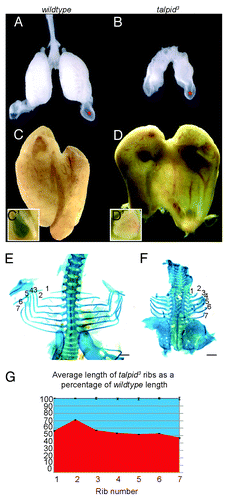
Figure 1. The talpid3 chicken exhibits abnormal lung and liver morphology. Compared with E10 wt lung (A), the talpid3 lung is smaller and poorly branched (B), air sac development was normal (red asterisk) (A andB). Wt liver (C) and talpid3 liver (D) are of similar size, although the talpid3 liver is green. The wt gall bladder is bile filled (green) (C’), while the talpid3 gall bladder lacks bile (D’). At E10 individual ribs were measured in wt (E) and compared with the corresponding rib in talpid3 (F). Average lengths were reduced in the talpid3 chicken; Rib one = 44% reduced, rib two = 28%, rib three = 44%, rib four = 46%, ribs five/six = 48%, rib seven = 53% smaller in talpid3 (G). Magnification is the same between A and B, C and D, and E and F.

Figure 2. Lung development in the wt and talpid3 chicken. Haematoxylin and eosin staining E7 (A–E). IHC anti-acetylated tubulin (cilia axonemes;green) and anti- γtubulin (centrosomes;red) E10 (CandF). At E7 the epithelial mesobronchi of each lung has branched extensively throughout the wt lung mesenchyme (asterisks) (A). The more mature wt primary bronchi (circled area) (A andB) are surrounded by a condensation of SeM cells (B). Mesobronchi branching in the talpid3 lung is disturbed and differs between lungs within the same embryo (bronchi labeled with asterisk or arrow asterisk) (D), the epithelia is thinner and disorganized and no SeM condensations are seen (E). E10 wt lung exhibit cilia (C) on epithelia (C’), SeM (C”) and SM cells (C”’). E10 talpid3 lung, cilia do not project from centrosomes (red box) (F and F’) Magnification comparable between A–E, C–F.
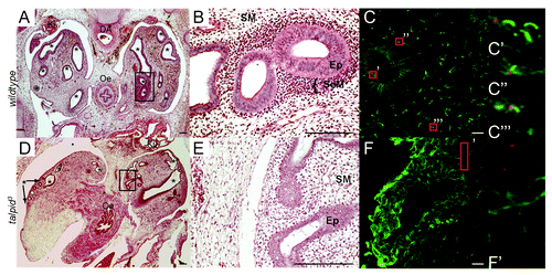
Figure 2. Lung development in the wt and talpid3 chicken. Haematoxylin and eosin staining E7 (A–E). IHC anti-acetylated tubulin (cilia axonemes;green) and anti- γtubulin (centrosomes;red) E10 (CandF). At E7 the epithelial mesobronchi of each lung has branched extensively throughout the wt lung mesenchyme (asterisks) (A). The more mature wt primary bronchi (circled area) (A andB) are surrounded by a condensation of SeM cells (B). Mesobronchi branching in the talpid3 lung is disturbed and differs between lungs within the same embryo (bronchi labeled with asterisk or arrow asterisk) (D), the epithelia is thinner and disorganized and no SeM condensations are seen (E). E10 wt lung exhibit cilia (C) on epithelia (C’), SeM (C”) and SM cells (C”’). E10 talpid3 lung, cilia do not project from centrosomes (red box) (F and F’) Magnification comparable between A–E, C–F.

Figure 3. Abnormal liver histology in the talpid3chicken. Haematoxylin and eosin staining E10 (A–F), E12 (G, H, J, K). IHC Cytokeratin19 (C’ andF’). Masson’s trichrome staining at E12 (IandL). E10, wt livers have large spaces throughout (AandB) and exhibit an immature version of the classic hepatic triumvirate with cholangiocytes arranged in a ring to produce early bile ducts (asterisk) (C) with bile (arrow) (C) but no Cytokeratin19 present (C’). E10 talpid3 liver is more compact, with fewer, smaller, spaces (D andE). Bile ducts develop but are overcrowded with cholangiocytes (F) which express Cytokeratin19 (F’) and bile is observed between the cholangiocytes (arrow) (F). E12 wt liver is more compact (G andH). E12 talpid3 liver presents areas of necrosis (J, K) and portal fibrosis (blue stain) (L) compared with wt (I).
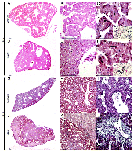
Figure 3. Abnormal liver histology in the talpid3chicken. Haematoxylin and eosin staining E10 (A–F), E12 (G, H, J, K). IHC Cytokeratin19 (C’ andF’). Masson’s trichrome staining at E12 (IandL). E10, wt livers have large spaces throughout (AandB) and exhibit an immature version of the classic hepatic triumvirate with cholangiocytes arranged in a ring to produce early bile ducts (asterisk) (C) with bile (arrow) (C) but no Cytokeratin19 present (C’). E10 talpid3 liver is more compact, with fewer, smaller, spaces (D andE). Bile ducts develop but are overcrowded with cholangiocytes (F) which express Cytokeratin19 (F’) and bile is observed between the cholangiocytes (arrow) (F). E12 wt liver is more compact (G andH). E12 talpid3 liver presents areas of necrosis (J, K) and portal fibrosis (blue stain) (L) compared with wt (I).

Figure 4. Hedgehog signaling is perturbed in the talpid3 liver during development. IHC anti-acetylated (cilia axonemes, green) and γtubulin (centrosomes, red) E6 (A andB). Wt liver has cilia axonemes projecting from centrosomes (circled) (A) cilia axonemes are not observed projecting from centrosomes in the talpid3 liver (circled) (B). (C) Real-time PCR identified a 0.08-fold reduction in PTCH1 expression in the day 6 talpid3 liver, compared with the wt.
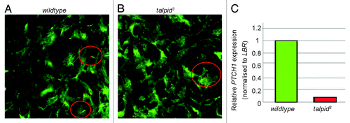
Figure 4. Hedgehog signaling is perturbed in the talpid3 liver during development. IHC anti-acetylated (cilia axonemes, green) and γtubulin (centrosomes, red) E6 (A andB). Wt liver has cilia axonemes projecting from centrosomes (circled) (A) cilia axonemes are not observed projecting from centrosomes in the talpid3 liver (circled) (B). (C) Real-time PCR identified a 0.08-fold reduction in PTCH1 expression in the day 6 talpid3 liver, compared with the wt.
Supplemental material

Additional material

Download Zip (8.7 MB)