23,501
Views
944
CrossRef citations to date
0
Altmetric
Review

Wnt signal transduction pathways

&
Pages 68-75 | Published online: 11 Jun 2008

Figures & data

Figure 1 A schematic representation of the canonical Wnt signal transduction cascade. Left, in the absence of Wnt ligand, a complex of Axin, APC, GSK3-β, CK1 and β-catenin is located in the cytosol. β-catenin is dually phosphorylated by CK1 and GSK3-β and targeted degraded by the proteosomal machinery mediated by β-TrCP. Right, with Wnt stimulation, signaling through the Fz receptor and LRP5/6 co-receptor complex induces the dual phosphorylation of LRP6 by CK1 and GSK3-β and this allows for the translocation of a protein complex containing Axin from the cytosol to the plasma membrane. Dsh is also recruited to the membrane and binds to Fz and Axin binds to phosphorylated LRP5/6. This complex formed at the membrane at Fz/LRP5/6 induces the stabilization of β-cat via either sequestration and/or degradation of Axin. β-catenin translocates into the nucleus where it complexes with Lef/Tcf family members to mediate transcriptional induction of target genes.
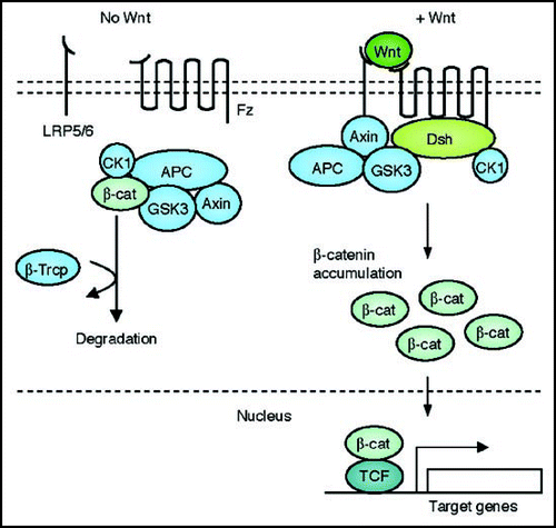
Figure 1 A schematic representation of the canonical Wnt signal transduction cascade. Left, in the absence of Wnt ligand, a complex of Axin, APC, GSK3-β, CK1 and β-catenin is located in the cytosol. β-catenin is dually phosphorylated by CK1 and GSK3-β and targeted degraded by the proteosomal machinery mediated by β-TrCP. Right, with Wnt stimulation, signaling through the Fz receptor and LRP5/6 co-receptor complex induces the dual phosphorylation of LRP6 by CK1 and GSK3-β and this allows for the translocation of a protein complex containing Axin from the cytosol to the plasma membrane. Dsh is also recruited to the membrane and binds to Fz and Axin binds to phosphorylated LRP5/6. This complex formed at the membrane at Fz/LRP5/6 induces the stabilization of β-cat via either sequestration and/or degradation of Axin. β-catenin translocates into the nucleus where it complexes with Lef/Tcf family members to mediate transcriptional induction of target genes.

Figure 2 A schematic representation of the Planar Cell Polarity transduction cascade. Wnt signaling is transduced through Fz independent of LRP5/6 leading to the activation of Dsh. Dsh through Daam1 mediates activation of Rho which in turn activates Rho kinase (ROCK). Daam1 also mediates actin polymerization through the actin binding protein Profilin. Dsh also mediates activation of Rac, which in turn activates JNK. The signaling from Rock, JNK and Profilin are integrated for cytoskeletal changes for cell polarization and motility during gastrulation.
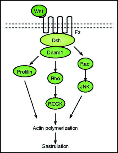
Figure 2 A schematic representation of the Planar Cell Polarity transduction cascade. Wnt signaling is transduced through Fz independent of LRP5/6 leading to the activation of Dsh. Dsh through Daam1 mediates activation of Rho which in turn activates Rho kinase (ROCK). Daam1 also mediates actin polymerization through the actin binding protein Profilin. Dsh also mediates activation of Rac, which in turn activates JNK. The signaling from Rock, JNK and Profilin are integrated for cytoskeletal changes for cell polarization and motility during gastrulation.

Figure 3 A schematic representation of the Wnt/Ca2+ signal transduction cascade. Wnt signaling via Fz mediates activation of Dsh via activation of G-proteins. Dishevelled activates the phosphodiesterase PDE which inhibits PKG and in turn inhibits Ca2+ release. Dsh through PLC activates IP3, which leads to release of intracellular Ca2+, which in turn activates CamK11 and calcineurin. Calcineurin activate NF-AT to regulate ventral cell fates. CamK11 activates TAK and NLK, which inhibit β-catenin/TCF function to negatively regulate dorsal axis formation. DAG through PKC activates CDC42 to mediate tissue separation and cell movements during gastrulation.
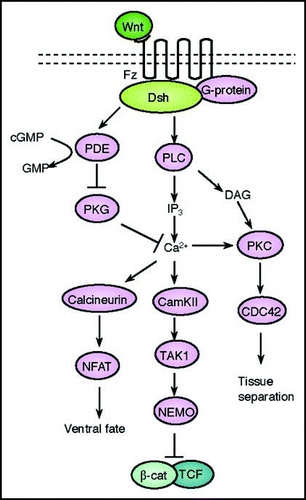
Figure 3 A schematic representation of the Wnt/Ca2+ signal transduction cascade. Wnt signaling via Fz mediates activation of Dsh via activation of G-proteins. Dishevelled activates the phosphodiesterase PDE which inhibits PKG and in turn inhibits Ca2+ release. Dsh through PLC activates IP3, which leads to release of intracellular Ca2+, which in turn activates CamK11 and calcineurin. Calcineurin activate NF-AT to regulate ventral cell fates. CamK11 activates TAK and NLK, which inhibit β-catenin/TCF function to negatively regulate dorsal axis formation. DAG through PKC activates CDC42 to mediate tissue separation and cell movements during gastrulation.

Reprints and Corporate Permissions

Please note: Selecting permissions does not provide access to the full text of the article, please see our help page How do I view content?

To request a reprint or corporate permissions for this article, please click on the relevant link below:

Academic Permissions

Please note: Selecting permissions does not provide access to the full text of the article, please see our help page How do I view content?

Obtain permissions instantly via Rightslink by clicking on the button below:

If you are unable to obtain permissions via Rightslink, please complete and submit this Permissions form. For more information, please visit our Permissions help page.