1,167
Views
48
CrossRef citations to date
0
Altmetric
Review

Wnt/β-catenin signaling in cerebral cortical development

Pages 76-80 | Published online: 11 Jun 2008

Figures & data

Figure 1 Expression of Wnts, Frizzled and Wnt activity in the developing cerebral cortex. Neural precursor cells reside in the ventricular zone (VZ). Ventricular cells are polarized neuroepithelial cells oriented with their apical surfaces towards the ventricular lumen; the endfeet of ventricular zone neural precursors are joined by β-catenin-rich adherens junctions (red). Wnt7a, Fz-5 and Fz-8 are expressed in the cortical ventricular zone, while Wnt7b is expressed in the developing cortical plate. Active Wnt/β-catenin signaling is present in the ventricular zone and cortical plate (blue). Increasing evidence points to important Wnt function in proliferation of precursors, laminar fate determination of cortical neurons, axonal/dendritic remodeling, as well as synapse formation.

Figure 1 Expression of Wnts, Frizzled and Wnt activity in the developing cerebral cortex. Neural precursor cells reside in the ventricular zone (VZ). Ventricular cells are polarized neuroepithelial cells oriented with their apical surfaces towards the ventricular lumen; the endfeet of ventricular zone neural precursors are joined by β-catenin-rich adherens junctions (red). Wnt7a, Fz-5 and Fz-8 are expressed in the cortical ventricular zone, while Wnt7b is expressed in the developing cortical plate. Active Wnt/β-catenin signaling is present in the ventricular zone and cortical plate (blue). Increasing evidence points to important Wnt function in proliferation of precursors, laminar fate determination of cortical neurons, axonal/dendritic remodeling, as well as synapse formation.

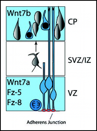
Table 1 CNS phenotype of Wnt knockouts