Figures & data
Figure 1 Experimental approaches. (A) Schematic drawing of a CA RS microscope. The inset window shows the energy diagram of CA RS. ωp: pump frequency; ωs: Stokes frequency; DM: dichroic mirror; PMT: photomultiplier tube. (B) Schematic of the composition of the gel scaffolds for the growth of DRG neurites. The figure is not to scale.
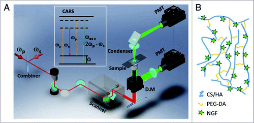
Figure 2 CA RS images of unstained live neurites growing from the DRG body cultured in 1% CS gel matrix. CA RS images of (A) live neurites at the proximity to the DRG body and (B) neurites and bulbs observed around the end of a neuronal growth cone. (C) Signal profile along a line indicated by the arrows in the inset image.
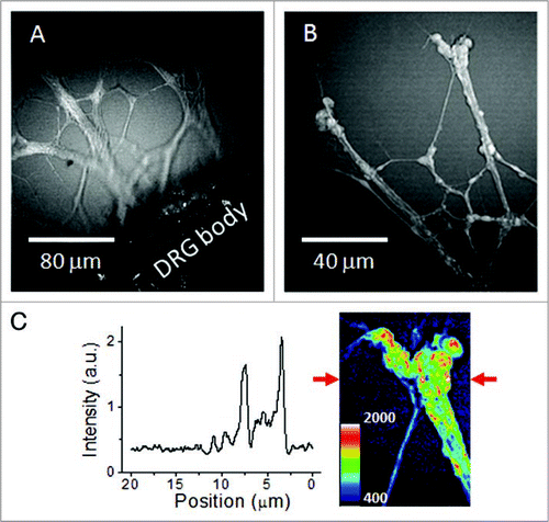
Figure 3 3-D structure of unstained neurites growing in a 1% CS gel matrix inspected by a laser-scanning CA RS microscope. Representative images at different depths show the 3-D distribution of the neurite growth. The number marked in each image indicates the depth relative to the bottom layer of neurite observed in the field of view.
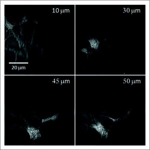
Figure 4 Monitoring growth of unstained neurites in hydrogels of 1% CS or 0.5% HA. (A–C) CA RS images of the neurite growth in a 1% CS matrix on day 1 to day 3. (D–E) images of the neurite growth in a 0.5% HA matrix on day 1 to day 3. The images are z-stacks of 15 µm in depth comprised of 16 optical sections. Arrows represent the direction of the neurite growth.
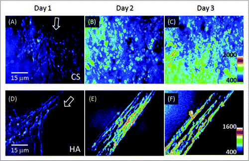
Figure 5 Analysis of CA RS signal of neurite growth: (A–C) In situ neurite growth in a 0.5% HA matrix on day 1 to day 3. (D) Relative density of neurites on day 1 through day 3. The analysis was performed according to the intensity in the 8-bit gray-scale 3-D images. Each data point represents the average number of the intensity results analyzed according to areas of three days (method).
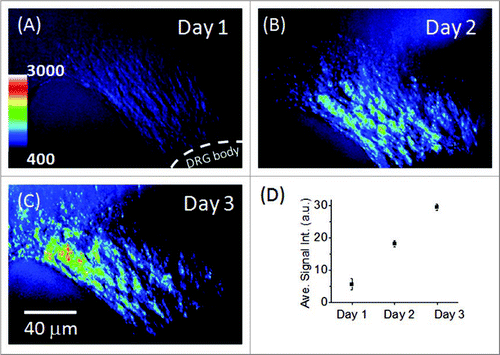
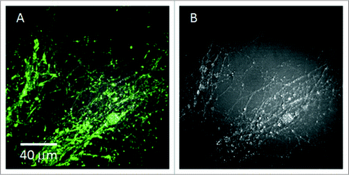
Figure 6 Phalloidin labeling of F-actin of neurites compared to CA RS images. (A) Overlaid image of signals from phalloidin labeling and CA RS. (B) CA RS image of the location shown in (A). The image exhibited lower contrast due to the higher non-resonant CARS signal resulting from the fixation of the sample.