Figures & data
Figure 1 Ex vivo angiogenesis in 3-dimensional matrigel cultures of fragments of the renal cortex or medulla obtained from 8-week-old and 22-week-old Zucker diabetic fat rats. Note that at the time of initiation of diabetes angiogenesis is enhanced, whereas at 22 weeks of age, when ECD is rampant, capillary sprouting is suppressed. These ex vivo findings were supported by the finding of a similar dynamics of microvascular density in the kidneys of these rats. Modified from ref. Citation40.
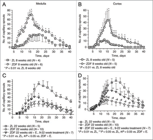
Figure 2 A schematic rendition of the concept of podokinesis—spontaneous or NO-stimulated turnover of focal adhesion complexes resulting in changes of tractional forces applied by the cells on the extracellular matrix. This is a scalar type of micromotion. This scalar motion is a prerequisite for acquisition of a directional movement when guidance cues are applied.
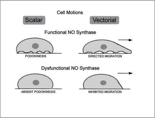
Figure 3 Cultured endothelial cells undergo endothelial mesenchymal transition induced by ADMA—loss of eNOS and acquisition of alpha-smooth muscle actin. Modified from ref. Citation70.
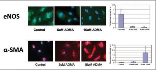
Figure 4 A hypothetical schema depicting the contribution of elevated guanidino compound, asymmetric dimethylarginine (ADMA), and downstream production of endostatin and TGFbeta to endothelial-mesenchymal transition, loss of vessel patency and fibrogenic transformation.
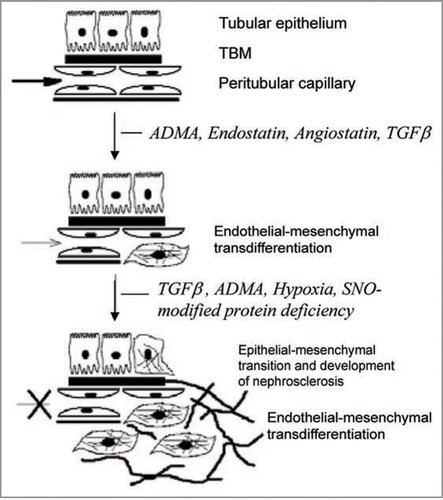
Figure 5 Mesenchymal stem cells are resident to many organs where they remain “sleepers” until stimulation, when cells may participate in angiogenesis, vasculogenesis and repair processes.
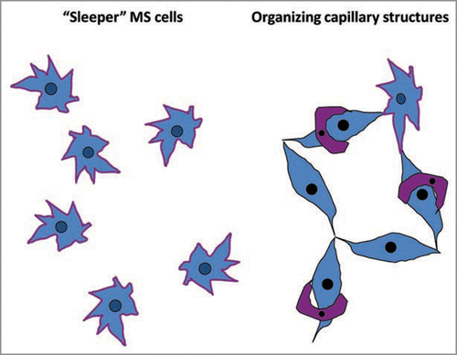
Figure 6 In patients with chronic allograft nephropathy, elevated excretion of endorepellin in the urine occurs at the expense of its parent molecule, perlecan, depletion of the latter from the glomerular and tubular basement membranes and diffusion of endorepellin into the renal parenchyma and the urine. Modified from ref. Citation86.
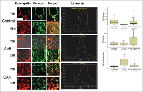
Table 1 Factors contributing to microvascular rarefaction