2,831
Views
35
CrossRef citations to date
0
Altmetric
Review

Palatogenesis

Engineering, pathways and pathologies

, , , , &
Pages 242-254 | Received 01 Jul 2011, Accepted 31 Aug 2011, Published online: 01 Oct 2011

Figures & data

Figure 1 Human palatal development. (A) Week six of human palatal development, with the secondary palate shown vertically on each side of the tongue and a gap between the secondary palate, nasal septum and primary palate. (B) After descent of the tongue, the secondary palatal shelves elevate and orient horizontally, allowing them to come in contact and begin fusing. (C) Fusion of the primary and secondary palate and the nasal septum separating the oropharynx from the nasopharynx. Figure modified from Dixon et al.Citation1
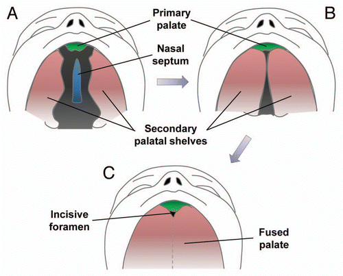
Figure 1 Human palatal development. (A) Week six of human palatal development, with the secondary palate shown vertically on each side of the tongue and a gap between the secondary palate, nasal septum and primary palate. (B) After descent of the tongue, the secondary palatal shelves elevate and orient horizontally, allowing them to come in contact and begin fusing. (C) Fusion of the primary and secondary palate and the nasal septum separating the oropharynx from the nasopharynx. Figure modified from Dixon et al.Citation1

Figure 2 Correlation between human and mouse palates. (A) Normal human upper lip, hard palate and soft palate. (B) Normal mouse upper lip, hard palate and soft palate. (C) Cleft of the secondary palate in human patient. (D) Clefting of secondary palate in transgenic mouse.
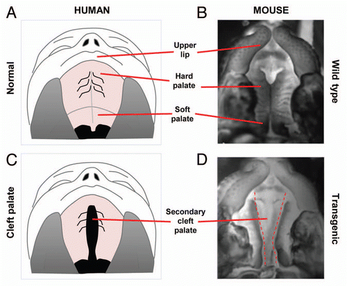
Figure 2 Correlation between human and mouse palates. (A) Normal human upper lip, hard palate and soft palate. (B) Normal mouse upper lip, hard palate and soft palate. (C) Cleft of the secondary palate in human patient. (D) Clefting of secondary palate in transgenic mouse.

Figure 3 Palate cultures. (A) Schematic of palate culture using 6-well plate and insert with 0.4 µM pores allowing cytokines but not cells to pass through. (B) Schematic of paired palatal shelves in palate culture. (C) H&E stain of palatal fusion after 72 h of 2 palatal shelves in culture while in contact with each other.
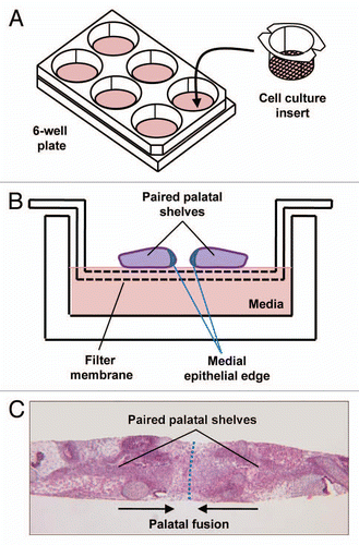
Figure 3 Palate cultures. (A) Schematic of palate culture using 6-well plate and insert with 0.4 µM pores allowing cytokines but not cells to pass through. (B) Schematic of paired palatal shelves in palate culture. (C) H&E stain of palatal fusion after 72 h of 2 palatal shelves in culture while in contact with each other.

Figure 4 Von Langenbeck palatal repair. (A) Secondary cleft palate palate with dotted red lines demonstrating incisions. (B) Mucuoperiosteal flap elevation with orange demonstrating opening of the incisions. (C) Midline nasal closure of the defect with the nasal layer in orange. (D) Closure of the midline incision with the oral mucosa over the nasal layer closure.
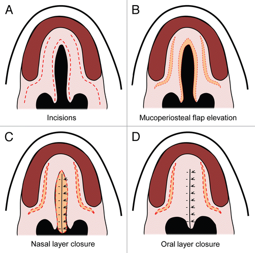
Figure 4 Von Langenbeck palatal repair. (A) Secondary cleft palate palate with dotted red lines demonstrating incisions. (B) Mucuoperiosteal flap elevation with orange demonstrating opening of the incisions. (C) Midline nasal closure of the defect with the nasal layer in orange. (D) Closure of the midline incision with the oral mucosa over the nasal layer closure.

Table 1 Genetic pathways involved in cleft lip and palate

Table 2 Summary of molecular pathways

Table 3 Summary of animal models used for palate study

Table 4 Procedure used for palate repair

Table 5 Summary of procedures used for palate repair

Table 6 Recent studies of palatal tissue engineering

Table 7 Summary of tissues described in tissue engineering of the palate

Table 8 Summary of animal models in studies discussing tissue engineering of the palate

Reprints and Corporate Permissions

Please note: Selecting permissions does not provide access to the full text of the article, please see our help page How do I view content?

To request a reprint or corporate permissions for this article, please click on the relevant link below:

Academic Permissions

Please note: Selecting permissions does not provide access to the full text of the article, please see our help page How do I view content?

Obtain permissions instantly via Rightslink by clicking on the button below:

If you are unable to obtain permissions via Rightslink, please complete and submit this Permissions form. For more information, please visit our Permissions help page.