Figures & data
Figure 1 Ureter maturation. (A and C) Whole mount and schematic of a mouse E11 urinary tract prior to ureter insertion. (B and D) Whole mount and schematic of an E14 urinary tract after ureter insertion. Note the ureter has now detached from the Wolffian duct and fused with the bladder epithelium.
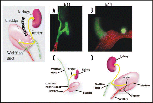
Figure 2 The Bladder Trigone. Top: Schematic of the ureteral valve mechanism and its physical relationship to the trigone. The trigone is a muscular structure located at the base of the bladder. When the bladder fills with urine, the trigonal muscle helps compress the ureteral orifice preventing back-flow of urine to the ureters and kidneys, which can cause severe damage. Bottom: The mouse ureteral valve. A vibratome section of a P0 urogenital tract stained with smooth muscle alpha actin (green) and uroplakin (red).
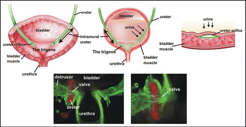
Figure 3 The Mackie Stephens hypothesis. Top: A ureteric bud emerging at the proper site on the Wolffian duct will join the trigone normally. Middle: A ureteric bud that forms too low on the Wolffian duct tends to join the bladder lateral or anterior to the normal insertion site causing reflux. Bottom: A ureteric bud forming too high on the Wolffian duct tends to remain attached to the sex ducts or joins the urogenital sinus posterior to its normal insertion site, causing obstruction. Green, Wolffian ducts and ureters; Red, cloacal endoderm and bladder trigone. Abbreviations: ub, ureteric bud; Wd, Wolffian duct.
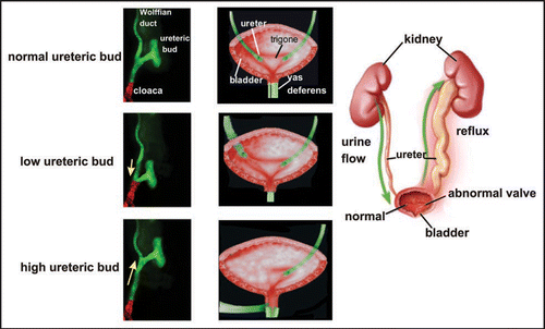
Figure 4 The original model of ureter insertion. Left: The ureter joins the bladder indirectly via the common nephric cut. Middle: The common nephric duct inserts into the bladder and expands, moving the ureter orifice anterior with respect to the Wolffian duct. Right: Further expansion of the trigone positions the ureter orifice in its final insertion site.
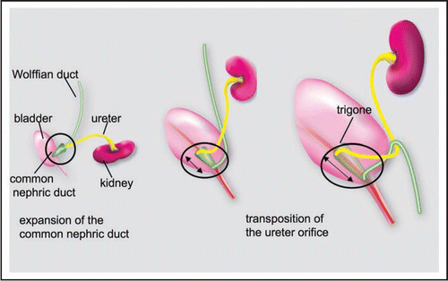
Figure 5 CND remodeling. (A) Left Top: Whole mount E12 Hoxb7-Gfp urogenital tract. Note that Gfp is present in Wolffian duct, ureter and CND but not in the bladder which is invisible in this image Bottom left: A sample of CND epithelium counterstained with E-cadherin showing the multilayered structure, as opposed to the simple tubular structure of the upper segments of Wolffian duct and ureter. Right: Images of Hoxb7-Gfp whole mount urogenital tracts between E11 and E14. Note the expansion of the CND (white dashed arrows at E11, E12 and E13) and its subsequent disappearance (E14, white dashed arrow). (B) Urogenital tracts from E11, E12 and E13 Hoxb7-Gfp embryos counterstained with cytokeratin (red) to illustrate expansion of the CND as the ureter moves into contact with the urogenital sinus. Abbreviations: CND, common nephric duct; ugs, urogenital sinus; Wd, Wolffian duct; ur, ureter.
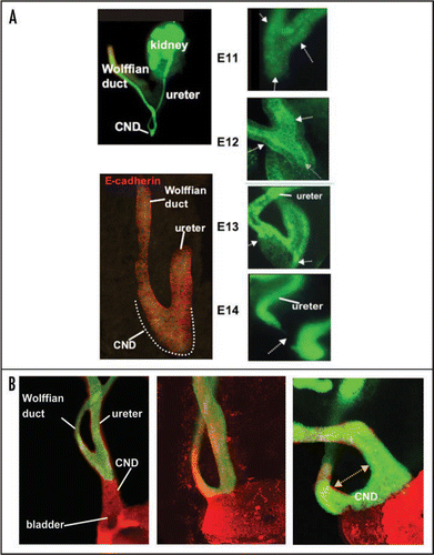
Figure 6 The CND does not form the trigone. Lineage analysis to determine the fate of CND cells. (A and D) A whole mount (A) and section (D) of a urogenital tract from an E13 Hoxb7-Cre;R26RlacZ embryo showing lacZ positive cells in the ureter, Wolffian duct and CND. (B and E) Whole mount (B) and section (E) of an E14 Hoxb7-Cre;R26RlacZ embryo showing lacZ expression at E14. Note the decline in lacZ expression in the CND but persistent expression in the ureters and Wolffian ducts. (C and F) Analysis of whole mount (C) and a section (F) of an urogenital tract from a E18 Hoxb7-Cre;R26RlacZ embryo. Note the absence of lacZ-labeled cells in the trigone, between the ureter and Wolffian duct where there is still strong lacZ expression. Bottom: A schematic showing a revised model of ureter maturation.
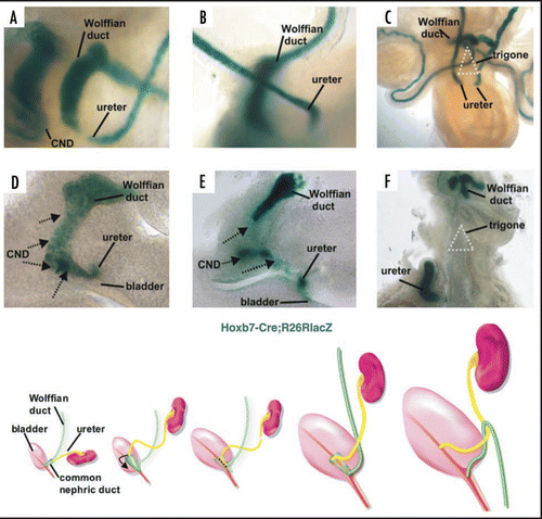
Figure 7 Apoptosis of the common nephric duct. Cryosection of am E11 Hoxb7-Gfp urogenital tract stained with activated caspase-3 antibody, (green) showing apoptosis of CND cells (red), but not cells in the Wolffian duct and ureter.
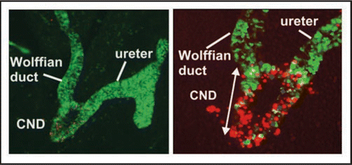
Figure 8 Fusion of the ureter orifice with the urogenital sinus and opening of the lumen. Left to right panels 1–6, Hoxb7-Gfp embryonic urogenital tracts stained with E-Cadherin (red) and activated caspase-3 (blue). Note apoptotic cells in the lumen of the ureter which is within the urogenital sinus epithelium at E15 (3,4) and the opening of the lumen on E16 (5,6).
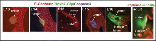
Figure 9 Ureter Insertion depends on alignment of the new ureter orifice with the bladder, regression of the CND, fusion of the ureter with the urogenital sinus epithelium and expansion of the bladder. Left: Vibratome sections of E12, E13 and E14 wild type urogenital tracts stained with antibody directed against cytokeratin (red).Note the enormous growth of the bladder relative to the urethra during this 3-day period. Right: Top, vibratome section of a Hoxb7-Gfp embryo (green) stained with E-cadherin (red). Note a loop has formed that helps align the ureter with the position in the urothelium (red) where it will fuse. Right middle and bottom: Laminin (red) stained Hoxb7-Gfp (green) urinary tracts showing the relative positions of the CND, ureter and Wolffian duct. At E13 the ureter orifice is fused with the urogenital sinus epithelium, but remains close to the Wolffian duct. Note the cell debris at this stage between the ureter and Wolffian duct where the CND was previously. Right Bottom: Between E13 and E14, the ureter orifice, which is tethered to the urogenital sinus epithelium, moves anteriorly as the bladder undergoes dramatic expansion.
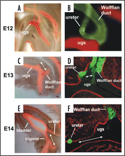
Figure 10 CND apoptosis depends on proximity to the bladder. (A and B) Control Hoxb7-Gfp urogenital tracts from E11 (A) and E18 (B) embryos. (C) Activated-Caspase-3 stained section of an E14 Hoxb7-Gfp embryo. (D) Ureter insertion in a P0 Hoxb7-Cre;R26RlacZ urogenital tract. (E and F) urogenital tracts from a Hoxb7-Gfp embryo exposed to retinoic acid; (E) E11, (F) E18. (G) Activated caspase-3 staining of the upper ureteric bud (ureteric bud 2) showing lack of apoptosis in the associated CND (CND2). (H) Ureter insertion in an E18 Hoxb7-Cre;R26RlacZ mouse with double ureters. Note the absence of CND1 and the persistence of the CND2.
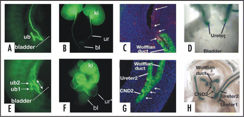
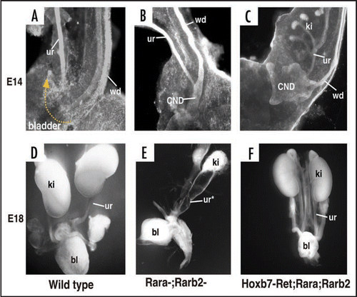
Figure 11 Defective CND remodeling in Rara-;Rarb2-mutants is rescued by Ret. (A) A cytokeratin stained E14 wild type urogenital tract. (B and C) Cytokeratin stained Rara-;Rarb2-mutant urogenital tract. (D) Whole mount E18 urogenital tract from a wild type embryo. (E) Whole mount E18 Rara-;Rarb2-urogenital tract. (F) Whole mount E18 Hoxb7-Cre;Rara-;Rarb2-urogenital tract.