661
Views
27
CrossRef citations to date
0
Altmetric
Review

Biological Roles of Prion Domains

, &
Pages 228-235 | Received 20 Dec 2008, Accepted 25 Feb 2008, Published online: 01 Oct 2007

Figures & data

Figure 1 Structural organization of prion proteins. QN: the QN-rich stretch. OR: the region of oligopeptide repeats. PrD-prion domain. Numbers correspond to amino acid (aa) positions. Arrows indicate domain and subdomain boundaries. N, M and C-N-proximal, middle and C-proximal regions of Sup35, respectively. The N/M and M/C boundaries are arbitrarily assigned to the second (aa 124) and third (aa 254) methionine residues of the Sup35 protein. See text for details.
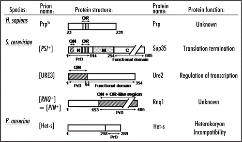
Figure 1 Structural organization of prion proteins. QN: the QN-rich stretch. OR: the region of oligopeptide repeats. PrD-prion domain. Numbers correspond to amino acid (aa) positions. Arrows indicate domain and subdomain boundaries. N, M and C-N-proximal, middle and C-proximal regions of Sup35, respectively. The N/M and M/C boundaries are arbitrarily assigned to the second (aa 124) and third (aa 254) methionine residues of the Sup35 protein. See text for details.

Figure 2 Evolutionary comparison of the N-terminal domains of Sup35 homologs. Sequences are from www.ncbi.nlm.nih.gov. Taxonomical relationships are from www.ncbi. nlm.nih.gov/Taxonomy. Scales do not correspond to evolutionary distances. For QN and OR designations, see . Numbers on the right correspond to the size of the N-terminal region (in aa) in each case. Sequence data were obtained from www.ncbi.nlm.nih.gov. ? -refers to the cases where search for prion activity in S. cerevisiae has been performed but have not yielded positive results (O. Zemlyanko, A. Petrova and G. Zhouravleva, unpublished; K. Gokhale and Y. Chernoff, unpublished). NT, not tested.
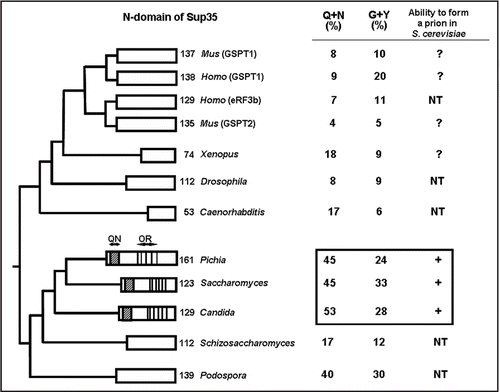
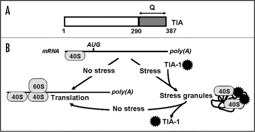
Figure 2 Evolutionary comparison of the N-terminal domains of Sup35 homologs. Sequences are from www.ncbi.nlm.nih.gov. Taxonomical relationships are from www.ncbi. nlm.nih.gov/Taxonomy. Scales do not correspond to evolutionary distances. For QN and OR designations, see Fig. 1. Numbers on the right correspond to the size of the N-terminal region (in aa) in each case. Sequence data were obtained from www.ncbi.nlm.nih.gov. ? -refers to the cases where search for prion activity in S. cerevisiae has been performed but have not yielded positive results (O. Zemlyanko, A. Petrova and G. Zhouravleva, unpublished; K. Gokhale and Y. Chernoff, unpublished). NT, not tested.
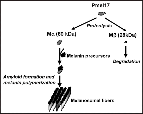
Figure 3 Formation of the stress granules. Schematic structure of TIA protein. (Q) the Q-rich stretch. Other designations are as in Model showing formation of stress granules. Ribosome subunits are shown as ovals and TIA as black asterisk. See text for more details.

Figure 3 Formation of the stress granules. Schematic structure of TIA protein. (Q) the Q-rich stretch. Other designations are as in Figure 1B Model showing formation of stress granules. Ribosome subunits are shown as ovals and TIA as black asterisk. See text for more details.

Figure 4 Role of amyloid in melanin polymerization. Glycoprotein Pmel17, that is a critical component of melanosome biogenesis, gives rise to two fragments, Mα and Mβ. Self-assembly of Mα leads to amyloid formation. Amyloid provides a scaffold for melanin polymerization.

Figure 4 Role of amyloid in melanin polymerization. Glycoprotein Pmel17, that is a critical component of melanosome biogenesis, gives rise to two fragments, Mα and Mβ. Self-assembly of Mα leads to amyloid formation. Amyloid provides a scaffold for melanin polymerization.

Reprints and Corporate Permissions

Please note: Selecting permissions does not provide access to the full text of the article, please see our help page How do I view content?

To request a reprint or corporate permissions for this article, please click on the relevant link below:

Academic Permissions

Please note: Selecting permissions does not provide access to the full text of the article, please see our help page How do I view content?

Obtain permissions instantly via Rightslink by clicking on the button below:

If you are unable to obtain permissions via Rightslink, please complete and submit this Permissions form. For more information, please visit our Permissions help page.