Figures & data
Table 1. Summary of the experimental design, attack rates (AR), survival times (ST) and global immunohistochemical results on the central nervous system (CNS), peripheral nervous system (PNS), gut-associated lymphoid tissues (GALT) and non-GALT (nGALT).
Figure 1. Influence of the host breed and genotype on the PrPd profiles when inoculated with ME7, 79A, 22A and 87V by the combined route. The x axis of the above graph refers to different morphological types of PrPd labeling identified by immunohistochemistry. PrPd types are: ITNR, intraneuronal; ITAS, intrastrocytic; ITMG, intramicroglial; STEL, stellate; SBPL, subpial; SBEP, subependymal; PRVS, perivascular; PVAC, perivacuolar; PART, fine particulate-coalescing; LINR, linear; PNER, perineuronal; EPEN, ependymal; NVPL, non vascular plaques; VASC, vascular plaques.
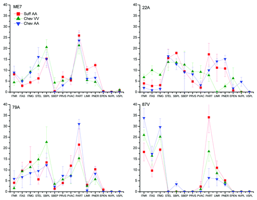
Table 2. Summary of the PrPd cell distribution according to strain and host.
Figure 2. Main PrPd features in brains of sheep infected with ME7, 79A, 22A, or 87V. In ME7 infections (A-C), neuropil-associated PrPd types predominated, like-wise perineuronal, coalescing or diffuse punctate. A prominent characteristic of the 22A agent in sheep (D-F) was the glia-associated PrPd types like the perivascular and perivacuolar in the white matter with some plaque-like depositions (arrows) in particular in Cheviots of the ARQ/ARQ genotype. 79A infections (G-I) resulted in a combination of intraglial-coalescing pattern (G) with stellate-like labeling (H, I). Sheep infected with 87V (J-L) unequivocally showed most PrPd associated with neurons or glial cells. Immunohistochemistry for PrPd using R145 antibody. Magnifications: x10 (D-F, H and I); x20 (A-C, G, J-L).
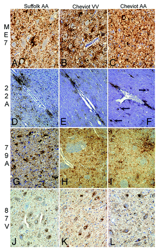
Figure 3. Epitope mapping results for the 87V infections in the dorsal motor nucleus of the vagus nerve of sheep. Note that Suffolk ARQ/ARQ sheep displayed intraneuronal immunolabelling with C-terminal antibodies such as Bar224 (A) and N-terminal antibodies such as P4 (B). Instead, Cheviot VRQ/VRQ (C, D) and ARQ/ARQ (E, F) sheep displayed the intraneuronal type with Bar224 (C, E) but not with P4 (D, F), a feature that is characteristic of strains like BSE and CH1641 when experimentally passaged in sheep. Immunohistochemistry for PrPd using Bar224 or P4 antibodies. Magnifications: x10 (A-D), x20 (E, F).
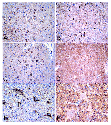
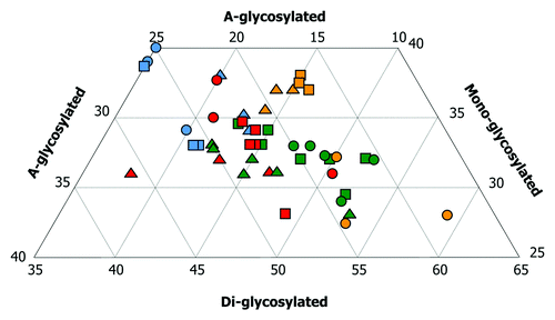
Figure 4. Triplot analyses from brains of sheep infected with cloned murine strains. Colors denote strain whereas symbol denotes breed and genotype. This plot shows total number of animals infected either by a single oral dose or a combined challenge. Eighteen sheep infected with ME7 (green) gave a positive result in Western-blot. Nine did with 79A (blue). Eleven sheep infected with 22A (red) gave a positive result in WB. All nine sheep infected with 87V (yellow) displayed strong signals of PrPres in their brains. Note that the PrPres associated with ovine ME7 showed higher amounts of the di-glycosylated band, followed by intermediate amounts of the mono-glycosylated band and low amounts of the a-glycosylated band. The characteristics of PrPres in ovine 22A infections are similar to ME7 ones but with a relatively lower and higher amount of the di- and a-glycosylated bands, respectively. The mono- and the a-glycosylated bands of PrPres are significantly higher after 79A infections. This pattern was similar to that observed for the PrPres associated with the 87V strain in Cheviot sheep of the VRQ/VRQ (triangles) and ARQ/ARQ (squares) genotypes, but not in the Suffolk sheep of the ARQ/ARQ genotype (circles), in which, the di-glycosylated band predominated.