Figures & data
Figure 1 Centrosome structure. Pericentriolar material surrounds a pair of barrels-shaped centrioles. Minus ends of astral microtubules lie in pericentriolar matrix and plus ends extend outward. The designated plus-end of microtubules is more dynamic, frequently growing or shrinking. The opposing minus end is also dynamic although tubulin is lost, but not added in vivo.
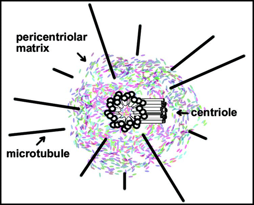
Figure 2 Spindle fusions in γ-tubulin mutants. Spindles in embryos from wild-type (wt) and γ-Tub37C (γ-mat) mutants as indicated. Embryos were stained with antibodies against α-tubulin to visualize microtubules and a fluorescent chromatin dye to visualize chromosomes. Arrows indicate some sites of apparent spindle fusion. Magnification identical in all panels.
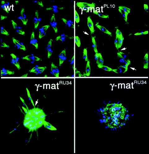
Figure 3 Centriole and basal body structure. Amorphous material is the first indication of a forming centriole or basal body. (A–C) The A-, B- and C-tubules appear sequentially. (D) Appendages form at the distal end of the parental centriole. (E–G) The transition zone marks the end of C-tubules in basal bodies. B- and C-tubules extend into axonemes. (H) The central pair of microtubules emerges in the transition zone, but they are not present in cilia. (E–H) The plasma membrane surrounding basal body/axonemes is depicted as a circle. Modified from tomography of centriole/basal body structure in Chlamydomonas.Citation33
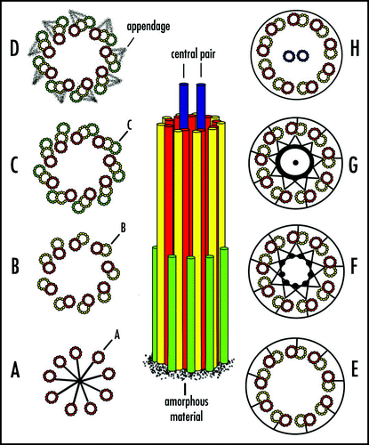
Figure 4 The centriole cycle and the spindle assembly checkpoint. (A) Normal centriole cycle. (B1) Failure in centriole duplication, but centrosomes with a single centriole assemble a bipolar spindle. (B2) A single centrosome is partitioned to daughter cells that generate a monopolar spindle. Centriole pairs are connected by fibrillar material, designated by black dots. Parental centriole shows distal appendages (blue lines) that may be vestiges of centriole assembly since they are not essential for mitosis or viability.Citation37 Pericentriolar matrix and microtubule asters are not depicted for simplicity.

Figure 5 Centriole checkpoint at the G1/S transition. Acentriolar cells were generated by microsurgery with the sharpened tip of glass pipetteCitation56 as shown here or by laser ablation.Citation57 Karyoplasts complete mitosis and often cytokinesis, arresting before passing the G1/S phase transition. Mitosis in control cells is depicted at the top. For simplicity, asters of microtubules in interphase cells are not shown.
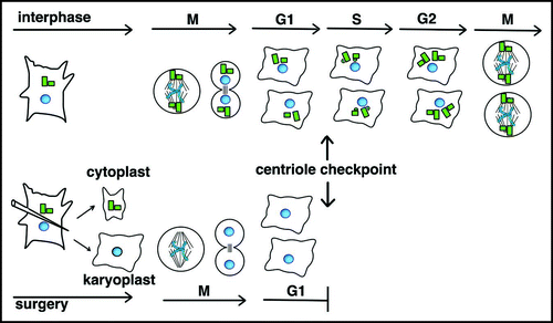
Figure 6 De novo assembly of centrioles. Following laser ablation of centrioles and completion of mitosis (M0), centrin-GFP in presumptive procentrioles (small dots) form in the first S-phase (S1). Pro/centrioles coalesce in G12 when centrioles acquire a robust pericentriolar mass and an aster of microtubules. Note that immature de novo assembled centrioles are depicted at the poles of a bipolar spindle at M1 for simplicity, but multipolar spindles were also formed. Because immature centrioles are not associated with microtubules, positioning of immature de novo assembled centrioles at spindle poles in M1 may reflect a microtubule-independent mechanism. Control cells retaining centrioles undergo normal cell cycle progression, but only G11-M1 is depicted here.
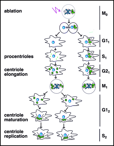
Table 1 γ-tubulin containing complexesTable Footnotea
Table 2 The extended tubulin superfamilyTable Footnotea