Figures & data
Figure 1 2D quantitative analysis of serum ceruloplasmin isoforms at 135 and 115 kDa. A master map was created utilizing PDQuest software from a triplicate of AD sample and a quadruplicate of control sample. The optical density analysis of the spots did not show statistically significant differences between AD and control samples.
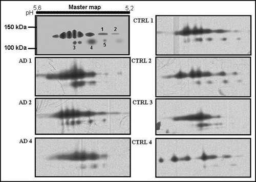
Figure 2 2D analysis of serum ceruloplasmin in AD (A) and controls (B). The pH 5–5.7 range is shown. Note the protein pattern similarity at about 135 and 115 kDa. Low-molecular-weight (<50 kDa) positive spots are present only in AD serum (A). 450 µl of a 1.5 mg/mL protein solution were loaded per gel. Ceruloplasmin content in both pools was brought 28 mg/dL and each serum sample contributed equally to the ceruloplasmin content.
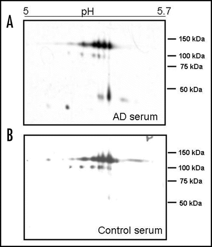
Figure 3 Box plot of the densitometric analysis (optical density percentage: % OD) of the ceruloplasmin spots immunoreactivity in controls and AD serum pools. The % ODs of the single spots obtained after the analysis of 2D PAGE films were analysed as a single “band” on the basis of their molecular weight corresponding to the 135 kDa, 115 kDa and <50 kDa forms. The sum of the 135 kDa % OD, 115 kDa % OD, 50 kDa % OD represents 100% of the ceruloplasmin immunoreactivity of a sample, which corresponds to 28 mg/dL of protein (see Methods). Black line displays the median and the box 90% of value distribution of four Western blots replicates performed in three sets of experiments. *p < 0.05.
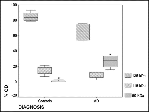
Table 1 Levels of serum copper and ceruloplasmin in controls and AD patients