Figures & data
Figure 1 Western blotting analysis of PrPSc from C-BSE and BSE/JP24. (A) The fragment of PrPSc in the cattle brain of C-BSE (lanes 1 and 3) and BSE/JP24 (lanes 2 and 4). PrPSc in the brain of TgBoPrP inoculated with C-BSE prion (lanes 5 and 7) and BSE/JP24 prion (lanes 6 and 8). All samples were digested with 50 µg/ml PK at 37°C for 1 h, and then samples in lanes 3, 4, 7 and 8 were treated with PNGaseF. PrPSc was detected by using mAb 6H4. Molecular markers are shown on the left (kDa). (B) The relative amount of the di-, mono- and non-glycosylated form of PrPSc in the C-BSE and BSE/JP24 prion affected individual. The results are mean ± standard deviation in five experiments. Bar diagram: diglycosylated form (black), monoglycosylated form (grey) and nonglycosylated form (white).
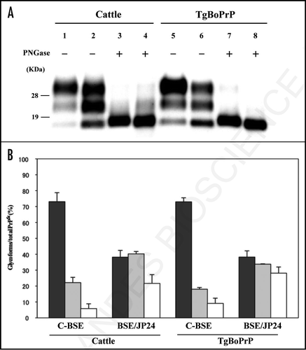
Figure 2 Lesion profile of TgBoPrP mice inoculated with C-BSE and BSE/JP24 prions. Vacuolation was scored on a 0–5 (mean values) in the following brain areas: 1, dorsal medulla; 2, cerebellar cortex; 3, superior cortex; 4, hypothalamus; 5, thalamus; 6, hippocampus; 7, septal nuclei of the paraterminal body; 8, cerebral cortex at the levels of 4 and 5; and 9, cerebral cortex at the level of 7. •: 1st passage of BSE/JP24 prion, ○: 2nd passage of BSE/JP24 prion, ▴: 1st passage of C-BSE prion.
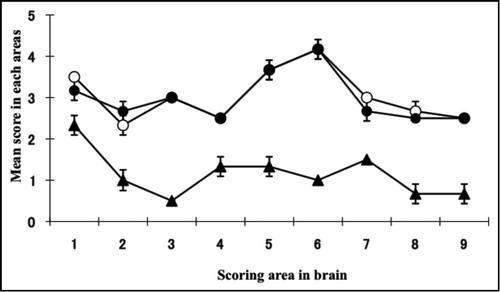
Figure 3 Histopathological (A and B), immunohistochemical (C and D) and PET-blot (E and F) analysis of TgBoPrP mice inoculated with C-BSE and BSE/JP24 prions. No distinct vacuolation in the presence of PrP plaques was detected in the cerebral cortex and hippocampal region in C-BSE prion affected TgBoPrP mice (A), whereas severe vacuolation in the absence of PrP-positive deposits was prominent in BSE/JP24 prion affected TgBoPrP mice (B). Immunolabelled PrPSc showed coarse granular and coalescing-like patterns in the gigantocellular nucleus of medulla oblongata of C-BSE prion affected TgBoPrP mice (C). A diffused fine granular pattern was observed in BSE/JP24 prion affected TgBoPrP mice (D). Immunohistochemical labelling with mAb F99/97.6.1. The insets at the lower right corner (A and C) are PrP plaques detected in the periventricular area of frontal lobe (arrowheads). PET blot reveals that immunolabelled PrPSc was marked in particular nuclei of brainstems in C-BSE prion affected TgBoPrP mice (E). On the other hand, widespread and homogeneous PrPSc immunolabelling was obvious in BSE/JP24 prion affected TgBoPrP mice (F). PET blots of the representative coronal section at the level of the hippocampus (left) and medulla oblongate (right) are shown. The mAb SAF84 was used in PET-blot analysis. Bar: 200 µm (A and B); 50 µm (C and D); 20 µm (inset of A and C).
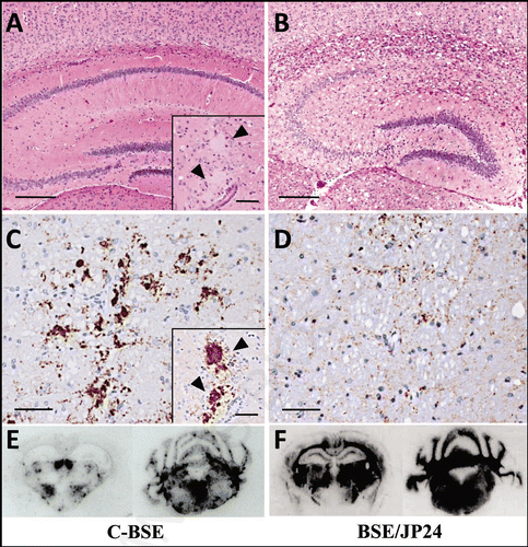
Figure 4 Relative PK resistance of PrPSc in prion affected cattle and TgBoPrP mice. (A) PK resistance of PrPSc in the cattle brain of C-BSE and BSE/JP24. (B) PK resistance of PrPSc of TgBoPrP mice inoculated with C-BSE and BSE/JP24 prions. (C) PK resistance of PrPSc from the subsequent passage of C-BSE and BSE/JP24 prion to TgBoPrP mice. The PrPSc concentration of sample was adjusted by the signal intensity of western blotting. The samples were treated with PK of various concentration (40–1,000 µg/ml) at 37°C for 1 h. Data shown represent one of three experiments demonstrating similar trends. PrPSc was detected with mAb 6H4. Molecular markers are shown on the left (kDa).
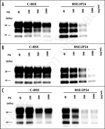
Table 1 Transmission of C-BSE and BSE/JP24 prion to mice
Table 2 Conformational stability ([GdnHCl]1/2 value) of PrPSc from C-BSE and BSE/JP24