Figures & data
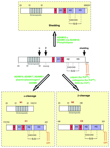
Figure 1. Schematic diagram of PrP processing. PrP is known to be cleaved at 3 sites: after residue 110 or 111 (α-cleavage), near the end of the octapeptide repeats region (β-cleavage), and at or near the GPI anchor (shedding). The amino acid numbering is based on human PrP. CHO: Asn-linked glycans; -S-S-: disulfide bridge; ROS: reactive oxygen species. The enzymes/factors involved in the processing are highlighted in green, and question marks denote the existence of conflicting reports on the respective protease(s).