Figures & data
Figure 1. Overall brain tractography from a 62 y-old male Parkinson patient (A) and a 61 y-old male healthy control (B). The color code represents fiber direction: blue-inferior to superior, red - left to right, green- anterior to posterior. The 3D view from the superior plane shows a diffuse reduction of white matter fibers in the PD patient compared with the control.
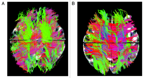
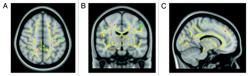
Figure 2. TBSS results and SPM-VBM analysis of cortical brain volume from 15 RR-MS patients and 15 healthy controls displayed on the Montreal Neurological Institute template. Clusters of reduced FA in patients compared with healthy controls are indicated in red. Areas of significant reduction of gray matter volume in patients compared with healthy controls are indicated in green.