Figures & data
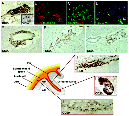
Figure 1.Classification of GM lesions as described by Bö et al.Citation8 Type I lesions (A) are GM/WM mixed lesions (original magnification 2.5x). The black line indicates the border between WM and GM. The GM and WM parts of the lesion are indicated with an arrow and arrowhead, respectively. The red arrowhead indicates a WM lesion. Type II lesions (B) are located completely intracortically, while type III and IV lesions run from the pial surface toward deeper cortical layers (D and E) or all the way to the GM/WM border, respectively (C; original magnification 5x). Both D en E are sections showing the same type III lesion. The lesion is visible in the MBP staining (D), showing a clearly demyelinated area (arrows indicate lesion borders), while the same area is invisible in a consecutive slide stained with a more conventional histochemical staining (E; Luxol Fast-Blue). The original magnification is 5x. (A-D), sections stained using myelin basic protein (MBP) antibodies. (E) Section stained using LFB.
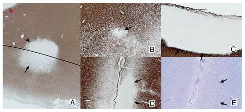
Table 1. topology of GM pathology in MS
Figure 2. Post-mortem MRI and accompanying PLP immunohistochemical staining of a coronal section. T2 weighted MRI scans (B) were compared with PLP stainings (A) of the same coronal sections. This figure clearly shows the difficulty to detect cortical lesions with conventional MRI sequences. (A): MRI-visible cortical lesions are shown in red, while MRI-invisible cortical lesions are shown in blue (original magnification 0.7x); (B1): MRI-visible lesions are indicated with arrowheads. MRI-visible cortical lesions show only very subtle increase of signal intensity; (B2) MRI-invisible lesions. Reproduced from Seewann et al.,Citation25 with permission from SAGE Publishers.
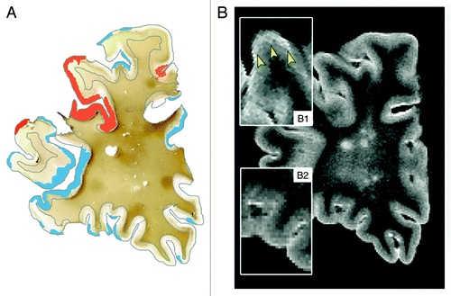
Figure 3. Meningeal inflammation in MS. Immunostainings of MS brain sections show intrameningeal ectopic B-cell follicle like structures in a cerebral sulcus containing CD20+ B cells (A), ramified stromal cells/FCD expressing CD35 (inset A) and expressing CXCLI3 (B), Ki67+ proliferating B cells (C), and plasmablast/plasma cells stained with an anti- Ig-G, -A, -M polyclonal antibody (D; the inset shows two intrafollicular plasma cells at high magnification). CD20+ B cells are also present in periventricular WM lesions (E) and in scarcely inflamed meninges entering a cerebral sulcus in a SPMS (F) and PPMS case (G). The lower schematic picture illustrates the localization of ectopic B-cell follicle like structures in the MS brain. These follicle like structures are present along (H) and in the depth (I) of the cerebral sulci, whereas scattered B lymphocytes (J) are detected in the meninges covering the external brain surface. Original magnifications: E-G = 100x; A, D, H-J = 200x, B, C and insets in A and D = 400x. Reproduced from Magliozzi et al.Citation59 with permission from Oxford University Press.