677
Views
4
CrossRef citations to date
0
Altmetric
Extra View

Not on the menu

Autophagy-independent clearance of prions

&
Pages 286-290 | Received 25 Jun 2013, Accepted 18 Jul 2013, Published online: 19 Jul 2013

Figures & data

Figure 1. Schematic presentation of PrP trafficking in infected cells and upon OHT treatment. In infected cells PrPC and PrPSc interact at the plasma membrane in cholesterol-rich lipid domains called lipid rafts. Upon internalization, both PrPC and PrPSc can recycle via the endosomal recycling compartment (ERC) or can be routed for degradation in lysosomes. Subcellular cholesterol distribution influences PrPSc trafficking in the endocytic pathway. In untreated, infected cells, the majority of PrP recycles through the cholesterol-rich ERC, supporting conversion of PrPC to PrPSc. Treatment with 4- hydroxytamoxifen (OHT) induces cholesterol accumulation in enlarged late endosomes (LE). PrPSc production and degradation defines the cellular load of infectious prions. We propose that 4-hydroxytamoxifen-induced changes in PrPSc trafficking favor PrPSc degradation. EE, early endosomes; LYS, lysosomes. (Adapted with permission from Marzo et al., 2013).Citation14
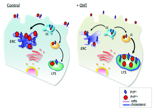
Figure 1. Schematic presentation of PrP trafficking in infected cells and upon OHT treatment. In infected cells PrPC and PrPSc interact at the plasma membrane in cholesterol-rich lipid domains called lipid rafts. Upon internalization, both PrPC and PrPSc can recycle via the endosomal recycling compartment (ERC) or can be routed for degradation in lysosomes. Subcellular cholesterol distribution influences PrPSc trafficking in the endocytic pathway. In untreated, infected cells, the majority of PrP recycles through the cholesterol-rich ERC, supporting conversion of PrPC to PrPSc. Treatment with 4- hydroxytamoxifen (OHT) induces cholesterol accumulation in enlarged late endosomes (LE). PrPSc production and degradation defines the cellular load of infectious prions. We propose that 4-hydroxytamoxifen-induced changes in PrPSc trafficking favor PrPSc degradation. EE, early endosomes; LYS, lysosomes. (Adapted with permission from Marzo et al., 2013).Citation14

Figure 2. Possible mechanisms of prion clearance by tamoxifen and OHT. Above dashed line: Tamoxifen and OHT may impair trafficking of cholesterol out of lysosomes. The high cholesterol environment favors PrPSc degradation and/or impairs PrPSc transconformation from PrPC substrate molecules (left lysosome). In contrast, without drug treatment normal cholesterol trafficking supports an environment that prevents PrPSc degradation and/or favors PrPSc formation (right lysosome). Below dashed line: Tamoxifen and OHT inhibit cholesterol biosynthesis at different steps. While tamoxifen treatment leads to zymosterol accumulation, OHT leads to desmosterol accumulation.Citation20 Desmosterol may be a more potent inhibitor of PrPSc formation or promoter of its degradation than zymosterol as indicated by the differently weighted lines impacting on PrPSc formation.
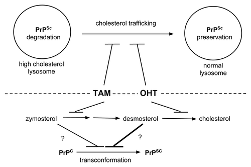
Figure 2. Possible mechanisms of prion clearance by tamoxifen and OHT. Above dashed line: Tamoxifen and OHT may impair trafficking of cholesterol out of lysosomes. The high cholesterol environment favors PrPSc degradation and/or impairs PrPSc transconformation from PrPC substrate molecules (left lysosome). In contrast, without drug treatment normal cholesterol trafficking supports an environment that prevents PrPSc degradation and/or favors PrPSc formation (right lysosome). Below dashed line: Tamoxifen and OHT inhibit cholesterol biosynthesis at different steps. While tamoxifen treatment leads to zymosterol accumulation, OHT leads to desmosterol accumulation.Citation20 Desmosterol may be a more potent inhibitor of PrPSc formation or promoter of its degradation than zymosterol as indicated by the differently weighted lines impacting on PrPSc formation.

Reprints and Corporate Permissions

Please note: Selecting permissions does not provide access to the full text of the article, please see our help page How do I view content?

To request a reprint or corporate permissions for this article, please click on the relevant link below:

Academic Permissions

Please note: Selecting permissions does not provide access to the full text of the article, please see our help page How do I view content?

Obtain permissions instantly via Rightslink by clicking on the button below:

If you are unable to obtain permissions via Rightslink, please complete and submit this Permissions form. For more information, please visit our Permissions help page.