Figures & data
Figure 1. Confocal image of an astrocyte transduced with NM-GFP encoding lentivirus. The cells were treated with 1 µM recombinant NM fibrils (monomer equivalent) for 24 h and fixed 48 h post fibril addition. A net-like NM-GFP aggregate structure with very high fluorescence intensity was visible close to the nucleus (left). Higher magnification of the cell periphery showed further spindle-shaped NM-GFP (green) aggregates present throughout the cytoplasm (right). Nuclei were stained with Hoechst (blue) and GFAP with anti-GFAP antibody (red). (Scale bar = 5 µm).
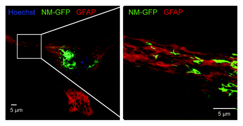
Figure 2. NM-HA aggregates transmitted from N2a donor to acceptor cells (Acc) co-localize with induced NM-GFP aggregates. Confocal microscopy revealed the presence of NM-HA and NM-GFP aggregates in the same N2a cell. Donor cells with induced NM-HA aggregates were co-cultured with acceptor cells expressing soluble NM-GFP. Different layers of a Z-stack are shown (Z distance = 0.564 µm). NM-HA aggregates were stained with anti-HA antibody (light blue) and F-actin was stained with iFluor-546-Phalloidin (red). Nuclei were stained with Hoechst (blue) and NM-GFP is displayed in green. (Scale bar = 5 μm).
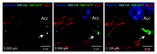
Figure 3. NM-HA aggregates in actin-rich protrusions. (A) Confocal image of a coculture of donor clone 2E (Do) and acceptor cells (Acc) expressing soluble NM-GFP. Cells were fixed after 48 h for immunofluorescence staining. The image shows one layer of a Z-stack. NM-HA aggregates of clone 2E were stained with anti-HA antibody (light blue) and F-actin was stained with iFluor-546-Phalloidin (red). The image shows transmitted light (gray) and NM-GFP in green. Nuclei were stained with Hoechst (blue). NM-GFP in the acceptor cell localizes in inclusions. The white square indicates the inset shown in B. (B) Excerpts of two layers of the Z-stack described above. The distance between the two layers is 1.830 μm. The distances to the bottom are indicated. The images show NM-HA aggregates in actin-rich protrusions between donor and acceptor cell. (Scale bar = 5 μm).
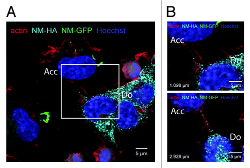
Figure 4. Life cycle of both PrP-derived and NM-derived prions in mammalian cells cultured ex vivo. Note that PrPC and NM differ in their cellular localization. PrPC is a cell surface bound glycoprotein, whereas NM is ectopically expressed in the cytosol. Both proteins are soluble and spontaneous conversion to a prion conformation has not been observed in mammalian cells ex vivo. The prion conformation can be induced by addition of infectious aggregates derived from the respective proteins. For PrP-derived prions, the inoculum is prepared from prion infected tissue or cell culture (for review, see ref. Citation4) or produced in vitro.Citation108 The prion phenotype is faithfully inherited by daughter cells during cell division. For PrP-derived prions, so far unknown susceptibility factors restrict persistent propagation to few cell lines (for review, see ref. Citation4). PrP-derived and NM-derived prions are transmitted horizontally to neighboring cells, in which they induce persistent aggregation of the prion proteins PrP or NM, respectively.
