Figures & data
Figure 1. (A) Cartoon representation of the X-ray crystal structure of ovine PrPC 128–233 (pdb 1UW3) with YYR in red and expanded SN6b epitope sequence highlighted in yellow (B) multiple sequence alignment of ovine, bovine, and murine PrP sequences (138–192, based on ovine sequence) with SN6b sequence highlighted as in panel A.
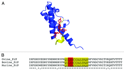
Table 1. SN6b-specific antibody titers for tga20 mice vaccinated at 0, 3, and 6 weeks with 10 μg of SN6b vaccine antigen (n = 6)
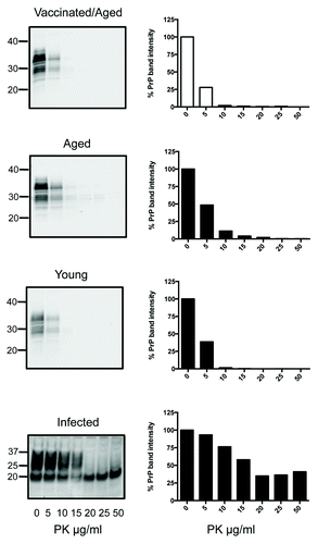
Figure 2. PK digests and western blots of combined brain homogenates from 6 SN6b-vaccinated, 4 age-matched, and 6 young tga20 mice. PK digests of an IP-infected (100 μl of 1% RML prion) mouse brain are shown for comparison. Error bars indicate mean and standard deviation of band intensities relative to samples not treated with PK for separate individual animal blots (not all blots shown).
Figure 3. PK digests and western blots of combined spleen homogenates from 6 SN6b-vaccinated, 4 age-matched, and 6 young tga20 mice. PK digests of the spleen from one IP infected (100 μl of 1% RML prion) tga20 mouse are shown for comparison (bottom panel).