719
Views
10
CrossRef citations to date
0
Altmetric
Research Paper

Homogenous photocatalytic decontamination of prion infected stainless steel and titanium surfaces

, , , , , & show all
Pages 488-495 | Received 22 May 2013, Accepted 12 Nov 2013, Published online: 18 Nov 2013

Figures & data

Figure 1. Photo-Fenton treatment of TiO2 (rutile) particles contaminated with the RML scrapie strain. 0.2 g of TiO2 were incubated with 10% w/v RML mouse brain homogenate and were then treated with 56 μg mL−1 Fe3+, 600 μg mL−1 h−1, UV-A, pH: 3.5. (A) SDS-PAGE, following AgNO3 staining of adsorbed proteins after treatment with photo-Fenton at the indicated time points (min). (B) western blotting of adsorbed PrP after treatment with photo-Fenton at the indicated time points (min). Primary antibody: 12F10, visualization: West Femto substrate (Pierce). Arrow: 32.5 kDa relative molecular mass marker.
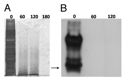
Figure 1. Photo-Fenton treatment of TiO2 (rutile) particles contaminated with the RML scrapie strain. 0.2 g of TiO2 were incubated with 10% w/v RML mouse brain homogenate and were then treated with 56 μg mL−1 Fe3+, 600 μg mL−1 h−1, UV-A, pH: 3.5. (A) SDS-PAGE, following AgNO3 staining of adsorbed proteins after treatment with photo-Fenton at the indicated time points (min). (B) western blotting of adsorbed PrP after treatment with photo-Fenton at the indicated time points (min). Primary antibody: 12F10, visualization: West Femto substrate (Pierce). Arrow: 32.5 kDa relative molecular mass marker.

Table 1. Efficiency of the following photo-Fenton decontamination of scrapie infected TiO2 particles based on a bioassay employing C57BL/6J mice

Figure 2. Photo-Fenton treatment of stainless steel and titanium wires contaminated with the 263K scrapie strain. Western blotting of PrP adsorbed on the surface of wires previously incubated with 10% w/v 263K hamster brain homogenate and then, treated with 56 μg mL−1 Fe3+, 300 μg mL−1 h−1 H2O2, UV-A, pH: 3.5, at the indicated time points (min). Primary antibody: 6Η4 (Prionics, AG), visualization: CDP-Star substrate (New England Biolabs). Arrow: 32.5 kDa relative molecular mass marker.
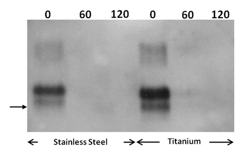
Figure 2. Photo-Fenton treatment of stainless steel and titanium wires contaminated with the 263K scrapie strain. Western blotting of PrP adsorbed on the surface of wires previously incubated with 10% w/v 263K hamster brain homogenate and then, treated with 56 μg mL−1 Fe3+, 300 μg mL−1 h−1 H2O2, UV-A, pH: 3.5, at the indicated time points (min). Primary antibody: 6Η4 (Prionics, AG), visualization: CDP-Star substrate (New England Biolabs). Arrow: 32.5 kDa relative molecular mass marker.

Figure 3. Scanning Electron Microscopy of stainless steel wires. a: uncontaminated, b-e: incubated with 10% hamster brain homogenate infected with the 263K prion strain and treated with 224 μg mL−1 Fe3+, 500 μg mL−1 h−1 H2O2, pH = 3.5, UV-A for 0, 240, 360, and 480 min respectively. White bar: 200μm, black bar: 20 μm. Panels to the right (A2-E2) are close ups of respective areas of the left column (A1-E1).
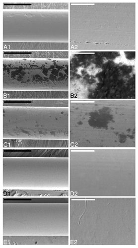
Figure 3. Scanning Electron Microscopy of stainless steel wires. a: uncontaminated, b-e: incubated with 10% hamster brain homogenate infected with the 263K prion strain and treated with 224 μg mL−1 Fe3+, 500 μg mL−1 h−1 H2O2, pH = 3.5, UV-A for 0, 240, 360, and 480 min respectively. White bar: 200μm, black bar: 20 μm. Panels to the right (A2-E2) are close ups of respective areas of the left column (A1-E1).

Figure 4. Fluorescence Microscopy of stainless steel wires. (A) uncontaminated, (B–E) incubated with 10% hamster brain homogenate infected with the 263K prion strain and treated with 224 μg mL−1 Fe3+, 500 μg mL−1 h−1 H2O2, pH = 3.5 and UV-A for 0, 240, 360, and 480 min respectively. Each row of panels (1–2) demonstrates areas of wires of the same group. Bar: 200 μm
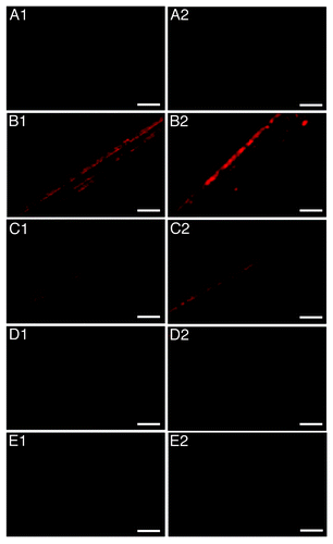
Figure 4. Fluorescence Microscopy of stainless steel wires. (A) uncontaminated, (B–E) incubated with 10% hamster brain homogenate infected with the 263K prion strain and treated with 224 μg mL−1 Fe3+, 500 μg mL−1 h−1 H2O2, pH = 3.5 and UV-A for 0, 240, 360, and 480 min respectively. Each row of panels (1–2) demonstrates areas of wires of the same group. Bar: 200 μm
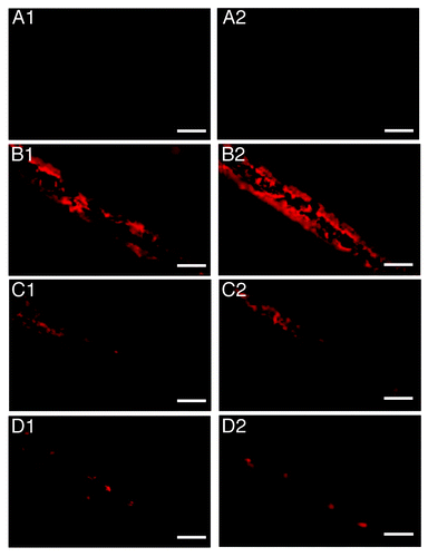
Figure 5. Fluorescence Microscopy of titanium wires. (A) uncontaminated, (B–D) incubated with 10% hamster brain homogenate infected with the 263K prion strain and treated with 224 μg mL−1 Fe3+, 500 μg mL−1 h−1 H2O2, pH = 3.5 and UV-A for 0, 240, and 480 min respectively. Each row of panels (1–2) demonstrates areas of wires of the same group. Bar: 200 μm

Figure 5. Fluorescence Microscopy of titanium wires. (A) uncontaminated, (B–D) incubated with 10% hamster brain homogenate infected with the 263K prion strain and treated with 224 μg mL−1 Fe3+, 500 μg mL−1 h−1 H2O2, pH = 3.5 and UV-A for 0, 240, and 480 min respectively. Each row of panels (1–2) demonstrates areas of wires of the same group. Bar: 200 μm

Figure 6. Surface protein contamination in groups of wires (n = 8), after densitometric analysis of fluorescence micrographs. (A) Stainless steel, (B) Titanium. Homogenous photocatalytic treatment was performed in the presence of 224 μg mL−1 Fe3+, 500 μg mL−1 h−1 H2O2, and UV-A (pH = 3.5).
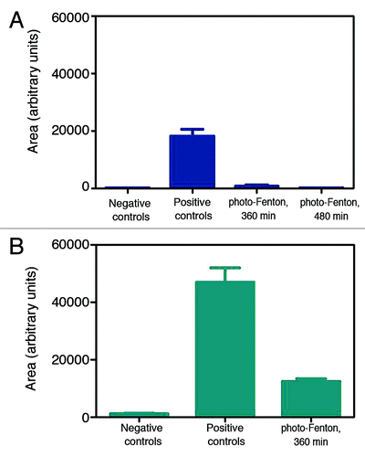
Figure 6. Surface protein contamination in groups of wires (n = 8), after densitometric analysis of fluorescence micrographs. (A) Stainless steel, (B) Titanium. Homogenous photocatalytic treatment was performed in the presence of 224 μg mL−1 Fe3+, 500 μg mL−1 h−1 H2O2, and UV-A (pH = 3.5).

Table 2. Bioassay of wires contaminated with the 263K scrapie strain

Supplemental material

Additional material

Download Zip (512.6 KB)

Reprints and Corporate Permissions

Please note: Selecting permissions does not provide access to the full text of the article, please see our help page How do I view content?

To request a reprint or corporate permissions for this article, please click on the relevant link below:

Academic Permissions

Please note: Selecting permissions does not provide access to the full text of the article, please see our help page How do I view content?

Obtain permissions instantly via Rightslink by clicking on the button below:

If you are unable to obtain permissions via Rightslink, please complete and submit this Permissions form. For more information, please visit our Permissions help page.