Figures & data
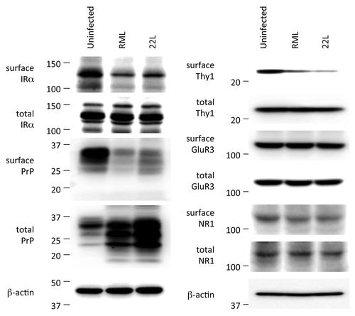
Figure 1. VSV-G(ts045)-GFP transport assay in uninfected N2aC24, infected N2aC24L1–3, and cured N2aC24L1–3 cells. Cells were transfected with the vector encoding VSV-G(ts045)-GFP and incubated at non-permissive temperature. Fluorescent intensities for VSV-G(ts045)-GFP at the Golgi region against those in the whole cell were determined in the randomly selected transfected cells (n = 14–16) at various times after the cells were transferred to permissive temperature. The Golgi area was immunohistochemically delineated using the Golgi marker GM130. Trafficking of VSV-G(ts045)-GFP in the cells was assessed as the accumulation kinetics of the protein in the Golgi area. Post-Golgi trafficking of VSV-G(ts045) was significantly delayed in infected N2aC24L1–3 cells, compared with that in uninfected N2aC24 cells. No significant difference in the VSV-G(ts045)-GFP trafficking was observed between uninfected N2aC24 and cured N2aC24L1–3 cells. Data were analyzed using the Student t test. * P < 0.05, ** P < 0.01. These were modified from Uchiyama et al.Citation8
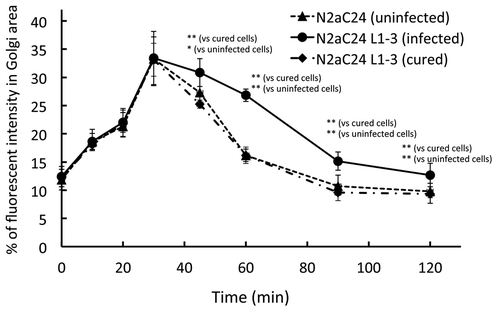
Figure 2. Western blotting of surface and total membrane proteins in the brains of uninfected and infected mice. Brains were freshly removed from uninfected control and terminally ill mice and coronally sliced. The brain slices were then subjected to biotin labeling of membrane proteins. The biotin-labeled surface proteins were purified using avidin-beads and subjected to western blotting for detection of IRα, PrP, Thy-1, GluR3, and NR1. Total expression levels of each protein were also determined. Biotinylated levels of IRα, PrP, and Thy-1, but not GluR3 and NR1, were lower in infected brains than in uninfected brains. Total expression levels of each protein were the same except for PrP. Total PrP was increased in infected brains due to accumulation of PrPSc. β-actin is an internal control. These were modified from Uchiyama et al.Citation8