Figures & data
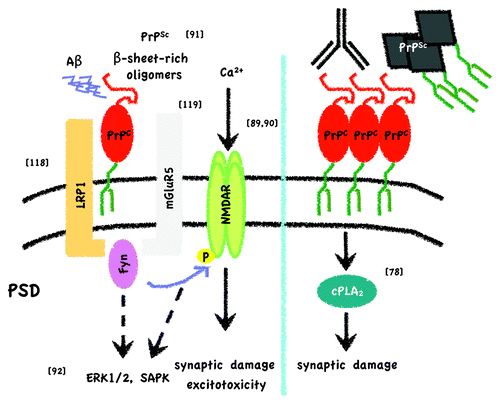
Figure 1. PrPC and PrPCGPIThy-1 expression in MDCK cells. (A) Confocal microscopy showing Z-stacks taken from top to bottom. As published beforeCitation33PrPC (in green) is basolaterally (b) sorted in fully polarized MDCK cells whereas changing the SS-GPI of PrPC for the one of Thy-1 preferentially directs the PrPCGPIThy-1 to the apical (a) compartment. Scale bar is 10μm. Originally published in PLoS One. (B) Schematic representation of the differential sorting of wild type (WT) PrPC and PrPCGPIThy-1 shown in A.
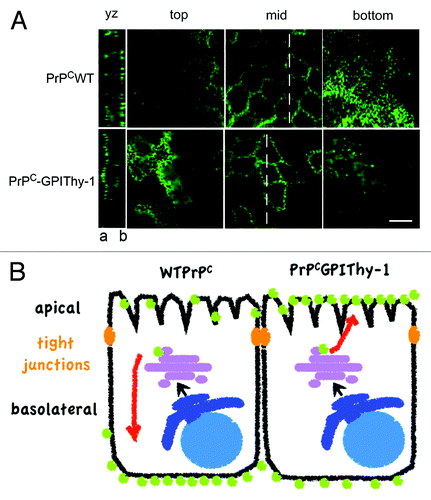
Figure 2. Simplified scheme of toxic signaling through PrPC. On the one hand (left) it has been described that PrPC can bind Aβ oligomers at the post-synaptic density (PSD), either derived from recombinant sources or from brains of Alzheimer disease patients, and elicit toxicity through Fyn activation, NMDAR phosphorylation and altered localization.Citation88,Citation90 Phosphorylation of NMDAR subunits would then lead to dendritic spine loss and excitotoxicty. As long as PrPC is attached to the outer leaflet of the membrane and Fyn to the inner, it has been proposed that a transmembrane protein should act as a scaffold. LRP1Citation118 and mGluR5Citation119 have been suggested as the interacting transmembrane partners after Aβ oligomer binding to PrPC. Resenberger et al. also showed that this signaling cascade can apply for various β-sheet rich conformers.Citation91 In the case of PrPSc binding, apart from Fyn activation, other signaling targets, such as ERK1/2, p38 and JNK, are seen to be activated in neuronal cell lines.Citation92 In addition, it might be hypothesized that β-sheet rich oligomer-associated NMDAR activation per se induces MAP kinase pathways. On the other hand (right) experiments by Bate and Williams demonstrated that after either crosslinking of PrPC with antibodies or by applying PrPSc to cortical primary neurons, there is an increase in membrane cholesterol content that leads to a toxic pathway implicating cPLA2 and arachidonic acid metabolites.Citation78 Reference to original studies is given by the numbers in brackets.